- PDB-3icf: Structure of Protein serine/threonine phosphatase from Saccharomy... -
+
Open data
ID or keywords:
Loading...
-
Basic information
Entry
Database: PDB / ID: 3icf
Title
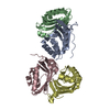
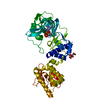
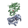
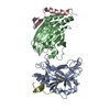
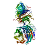
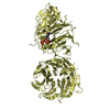
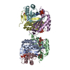
Structure of Protein serine/threonine phosphatase from Saccharomyces cerevisiae with similarity to human phosphatase PP5
Components
Serine/threonine-protein phosphatase T
Keywords
HYDROLASE / phosphatase / serine/threonine / saccharomyces cerevisiae / iron / metalloprotein / Structural Genomics / PSI-2 / Protein Structure Initiative / Midwest Center for Structural Genomics / MCSG / Manganese / Metal-binding / Nucleus / Protein phosphatase / TPR repeat
Function / homology
Function and homology information
Recruitment and ATM-mediated phosphorylation of repair and signaling proteins at DNA double strand breaks / protein-serine/threonine phosphatase / protein serine/threonine phosphatase activity / metal ion binding / nucleus / cytosol / cytoplasm Similarity search - Function
PP5, C-terminal metallophosphatase domain / PPP domain / PPP5 TPR repeat region / : / Serine/threonine specific protein phosphatases signature. / Protein phosphatase 2A homologues, catalytic domain. / Serine/threonine-specific protein phosphatase/bis(5-nucleosyl)-tetraphosphatase / Metallo-dependent phosphatases / Purple Acid Phosphatase; chain A, domain 2 / Calcineurin-like phosphoesterase domain, ApaH type ...PP5, C-terminal metallophosphatase domain / PPP domain / PPP5 TPR repeat region / : / Serine/threonine specific protein phosphatases signature. / Protein phosphatase 2A homologues, catalytic domain. / Serine/threonine-specific protein phosphatase/bis(5-nucleosyl)-tetraphosphatase / Metallo-dependent phosphatases / Purple Acid Phosphatase; chain A, domain 2 / Calcineurin-like phosphoesterase domain, ApaH type / Calcineurin-like phosphoesterase / Metallo-dependent phosphatase-like / TPR repeat region circular profile. / TPR repeat profile. / Tetratricopeptide repeats / Tetratricopeptide repeat / 4-Layer Sandwich / Tetratricopeptide-like helical domain superfamily / Alpha Beta Similarity search - Domain/homology
Mass: 18.015 Da / Num. of mol.: 76 / Source method: isolated from a natural source / Formula: H2O
-
Details
Has protein modification
Y
Sequence details
THE AUTHOR STATES THAT THE STRUCTURE WAS SOLVED WITH LIMITING AMOUNTS OF V8 PROTEASE ADDED TO THE ...THE AUTHOR STATES THAT THE STRUCTURE WAS SOLVED WITH LIMITING AMOUNTS OF V8 PROTEASE ADDED TO THE CRYSTALLIZATION LIQUOR AS A WAY TO ENHANCE CRYSTALLIZATION. THUS THOUGH THE INITIAL PROTEIN WAS INDEED FULL-LENGTH, ONLY THE CATALYTIC DOMAIN IS OBSERVED IN THE STRUCTURE, NOT THE N-TERMINAL TPR REPEATS. WE HAVE NOT CONFIRMED THE CRYSTALLIZED FRAGMENT BY MASS SPEC, BUT BELEIVE THE N-TERMINAL 178 RESIDUES HAVE BEEN REMOVED BY THE V8 PROTEASE.
-
Experimental details
-
Experiment
Experiment
Method: X-RAY DIFFRACTION / Number of used crystals: 1
-
Sample preparation
Crystal
Density Matthews: 2.04 Å3/Da / Density % sol: 39.62 %
Crystal grow
Temperature: 294 K / Method: vapor diffusion, sitting drop / pH: 7.5 Details: 0.1 M Hepes 7.5, 0.2 M NaCl, 25% PEG3350 plus 0.015 mg/ml V8 protease. Cryoprotected with Paratone-N oil, VAPOR DIFFUSION, SITTING DROP, temperature 294K
In the structure databanks used in Yorodumi, some data are registered as the other names, "COVID-19 virus" and "2019-nCoV". Here are the details of the virus and the list of structure data.
Jan 31, 2019. EMDB accession codes are about to change! (news from PDBe EMDB page)
EMDB accession codes are about to change! (news from PDBe EMDB page)
The allocation of 4 digits for EMDB accession codes will soon come to an end. Whilst these codes will remain in use, new EMDB accession codes will include an additional digit and will expand incrementally as the available range of codes is exhausted. The current 4-digit format prefixed with “EMD-” (i.e. EMD-XXXX) will advance to a 5-digit format (i.e. EMD-XXXXX), and so on. It is currently estimated that the 4-digit codes will be depleted around Spring 2019, at which point the 5-digit format will come into force.
The EM Navigator/Yorodumi systems omit the EMD- prefix.
Related info.:Q: What is EMD? / ID/Accession-code notation in Yorodumi/EM Navigator
Yorodumi is a browser for structure data from EMDB, PDB, SASBDB, etc.
This page is also the successor to EM Navigator detail page, and also detail information page/front-end page for Omokage search.
The word "yorodu" (or yorozu) is an old Japanese word meaning "ten thousand". "mi" (miru) is to see.
Related info.:EMDB / PDB / SASBDB / Comparison of 3 databanks / Yorodumi Search / Aug 31, 2016. New EM Navigator & Yorodumi / Yorodumi Papers / Jmol/JSmol / Function and homology information / Changes in new EM Navigator and Yorodumi
 Movie
Movie Controller

 Yorodumi
 Open data
 Basic information
 Components
 Keywords
 Function and homology information
 X-RAY DIFFRACTION /
  Authors
 Citation
 Structure visualization
 Downloads & links
 Other downloads
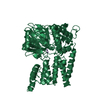


 PDBj
PDBj


 Assembly
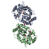


 Mass: 55.845 Da / Num. of mol.: 4 / Source method: obtained synthetically / Formula: Fe
 Mass: 94.971 Da / Num. of mol.: 2 / Source method: obtained synthetically / Formula: PO4
 Mass: 62.068 Da / Num. of mol.: 2 / Source method: obtained synthetically / Formula: C2H6O2
 Mass: 22.990 Da / Num. of mol.: 1 / Source method: obtained synthetically / Formula: Na
 Mass: 35.453 Da / Num. of mol.: 1 / Source method: obtained synthetically / Formula: Cl
 Sample preparation
 / Beamline: 19-ID / Wavelength: 0.97926 Å
 Processing