 Movie
Movie Controller

[English] 日本語
 Yorodumi
- PDB-3zm8: Crystal structure of Podospora anserina GH26-CBM35 beta-(1,4)- ma... -
+ Open data

- Basic information
| Entry | Database: PDB / ID: 3zm8 | |||||||||
|---|---|---|---|---|---|---|---|---|---|---|
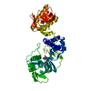
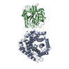
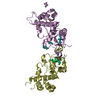
| Title | Crystal structure of Podospora anserina GH26-CBM35 beta-(1,4)- mannanase | |||||||||
 Components Components | GH26 ENDO-BETA-1,4-MANNANASE | |||||||||
 Keywords Keywords | HYDROLASE / GLYCOSYL HYDROLASE / CAZY / GH5 | |||||||||
| Function / homology |  Function and homology information Function and homology informationsubstituted mannan metabolic process / mannan endo-1,4-beta-mannosidase / mannan endo-1,4-beta-mannosidase activity / carbohydrate binding / metal ion binding Similarity search - Function | |||||||||
| Biological species |  PODOSPORA ANSERINA (fungus) PODOSPORA ANSERINA (fungus) | |||||||||
| Method |  X-RAY DIFFRACTION / SYNCHROTRON / MOLECULAR REPLACEMENT / Resolution: 2.85 Å X-RAY DIFFRACTION / SYNCHROTRON / MOLECULAR REPLACEMENT / Resolution: 2.85 Å | |||||||||
 Authors Authors | Couturier, M. / Roussel, A. / Rosengren, A. / Leone, P. / Stalbrand, H. / Berrin, J.G. | |||||||||
 Citation Citation | Journal: J.Biol.Chem. / Year: 2013 Title: Structural and Biochemical Analyses of Glycoside Hydrolase Families 5 and 26 Beta-(1,4)-Mannanases from Podospora Anserina Reveal Differences Upon Manno-Oligosaccharides Catalysis. Authors: Couturier, M. / Roussel, A. / Rosengren, A. / Leone, P. / Stalbrand, H. / Berrin, J.G. | |||||||||
| History |
| |||||||||
| Remark 700 | SHEET DETERMINATION METHOD: DSSP THE SHEETS PRESENTED AS "AE" IN EACH CHAIN ON SHEET RECORDS BELOW ... SHEET DETERMINATION METHOD: DSSP THE SHEETS PRESENTED AS "AE" IN EACH CHAIN ON SHEET RECORDS BELOW IS ACTUALLY AN 8-STRANDED BARREL THIS IS REPRESENTED BY A 9-STRANDED SHEET IN WHICH THE FIRST AND LAST STRANDS ARE IDENTICAL. |
- Structure visualization
| Structure viewer | Molecule: MolmilJmol/JSmol |
|---|
- Downloads & links
-Download
| PDBx/mmCIF format | 3zm8.cif.gz | 106.6 KB | Display | PDBx/mmCIF format |
|---|---|---|---|---|
| PDB format | pdb3zm8.ent.gz | 80.2 KB | Display | PDB format |
| PDBx/mmJSON format | 3zm8.json.gz | Tree view | PDBx/mmJSON format | |
| Others |  Other downloads Other downloads |
-Validation report
| Arichive directory | https://data.pdbj.org/pub/pdb/validation_reports/zm/3zm8ftp://data.pdbj.org/pub/pdb/validation_reports/zm/3zm8 | HTTPS FTP |
|---|
-Related structure data
| Related structure data |  3zizC  2bvtS 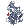 2qhaS  2vx4S 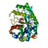 2whkS C: citing same article ( S: Starting model for refinement |
|---|---|
| Similar structure data |


-Links
 PDBj
PDBj


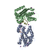
- Assembly
| Deposited unit | 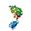
| ||||||||
|---|---|---|---|---|---|---|---|---|---|
| 1 |
| ||||||||
| Unit cell |
|
-Components
-Protein / Sugars , 2 types, 2 molecules A
| #1: Protein | Mass: 52848.484 Da / Num. of mol.: 1 Source method: isolated from a genetically manipulated source Source: (gene. exp.) PODOSPORA ANSERINA (fungus) / Strain: S MAT+ / Plasmid: PPICZAC / Production host: KOMAGATAELLA PASTORIS (fungus) / Strain (production host): X33References: UniProt: E2GHW2, mannan endo-1,4-beta-mannosidase |
|---|---|
| #2: Polysaccharide | 2-acetamido-2-deoxy-beta-D-glucopyranose-(1-4)-2-acetamido-2-deoxy-beta-D-glucopyranose Source method: isolated from a genetically manipulated source |
-Non-polymers , 4 types, 146 molecules 


| #3: Chemical | ChemComp-CA /  Mass: 40.078 Da / Num. of mol.: 1 / Source method: obtained synthetically / Formula: Ca Mass: 40.078 Da / Num. of mol.: 1 / Source method: obtained synthetically / Formula: Ca |
|---|---|
| #4: Chemical | ChemComp-HG /  Mass: 200.590 Da / Num. of mol.: 1 / Source method: obtained synthetically / Formula: Hg Mass: 200.590 Da / Num. of mol.: 1 / Source method: obtained synthetically / Formula: Hg |
| #5: Chemical | ChemComp-TLA /  Mass: 150.087 Da / Num. of mol.: 1 / Source method: obtained synthetically / Formula: C4H6O6 Mass: 150.087 Da / Num. of mol.: 1 / Source method: obtained synthetically / Formula: C4H6O6 |
| #6: Water | ChemComp-HOH / Mass: 18.015 Da / Num. of mol.: 143 / Source method: isolated from a natural source / Formula: H2O |
-Details
| Has protein modification | Y |
|---|
-Experimental details
-Experiment
| Experiment | Method: X-RAY DIFFRACTION / Number of used crystals: 1 |
|---|
- Sample preparation
| Crystal | Density Matthews: 2.32 Å3/Da / Density % sol: 47 % / Description: NONE |
|---|---|
| Crystal grow | pH: 7 Details: 0.1 M TRIS PH 7.0, 0.2 M NACL, 0.8 M POTASSIUM SODIUM TARTRATE, 1 MM HGCL2 |
-Data collection
| Diffraction | Mean temperature: 100 K |
|---|---|
| Diffraction source | Source: SYNCHROTRON / Site: ESRF  / Beamline: ID14-1 / Wavelength: 0.97914 / Beamline: ID14-1 / Wavelength: 0.97914 |
| Detector | Type: ADSC CCD / Detector: CCD / Date: Jun 7, 2012 |
| Radiation | Protocol: SINGLE WAVELENGTH / Monochromatic (M) / Laue (L): M / Scattering type: x-ray |
| Radiation wavelength | Wavelength: 0.97914 Å / Relative weight: 1 |
| Reflection | Resolution: 2.85→50 Å / Num. obs: 18575 / % possible obs: 99.9 % / Observed criterion σ(I): 2 / Redundancy: 11.8 % / Biso Wilson estimate: 85.54 Å2 / Rmerge(I) obs: 0.08 / Net I/σ(I): 25.6 |
| Reflection shell | Resolution: 2.85→3 Å / Redundancy: 11.8 % / Rmerge(I) obs: 0.65 / Mean I/σ(I) obs: 4.6 / % possible all: 100 |
- Processing
| Software |
| ||||||||||||||||||||||||||||||||||||||||||||||||||||||||||||||||||||||||||||||||||||||||||||||||||||||||||||||||||
|---|---|---|---|---|---|---|---|---|---|---|---|---|---|---|---|---|---|---|---|---|---|---|---|---|---|---|---|---|---|---|---|---|---|---|---|---|---|---|---|---|---|---|---|---|---|---|---|---|---|---|---|---|---|---|---|---|---|---|---|---|---|---|---|---|---|---|---|---|---|---|---|---|---|---|---|---|---|---|---|---|---|---|---|---|---|---|---|---|---|---|---|---|---|---|---|---|---|---|---|---|---|---|---|---|---|---|---|---|---|---|---|---|---|---|---|
| Refinement | Method to determine structure: MOLECULAR REPLACEMENT Starting model: PDB ENTRIES 2QHA, 2BVT, 2VX4 AND 2WHK Resolution: 2.85→28.3 Å / Cor.coef. Fo:Fc: 0.9267 / Cor.coef. Fo:Fc free: 0.8774 / SU R Cruickshank DPI: 0.58 / Cross valid method: THROUGHOUT / σ(F): 0 / SU R Blow DPI: 0.705 / SU Rfree Blow DPI: 0.333 / SU Rfree Cruickshank DPI: 0.327 Details: IDEAL-DIST CONTACT TERM CONTACT SETUP. RESIDUE TYPES WITHOUT CCP4 ATOM TYPE IN LIBRARY=CA HG. NUMBER OF ATOMS WITH PROPER CCP4 ATOM TYPE=3674. NUMBER WITH APPROX DEFAULT CCP4 ATOM TYPE=0. ...Details: IDEAL-DIST CONTACT TERM CONTACT SETUP. RESIDUE TYPES WITHOUT CCP4 ATOM TYPE IN LIBRARY=CA HG. NUMBER OF ATOMS WITH PROPER CCP4 ATOM TYPE=3674. NUMBER WITH APPROX DEFAULT CCP4 ATOM TYPE=0. NUMBER TREATED BY BAD NON-BONDED CONTACTS=2.
| ||||||||||||||||||||||||||||||||||||||||||||||||||||||||||||||||||||||||||||||||||||||||||||||||||||||||||||||||||
| Displacement parameters | Biso mean: 68.26 Å2
| ||||||||||||||||||||||||||||||||||||||||||||||||||||||||||||||||||||||||||||||||||||||||||||||||||||||||||||||||||
| Refine analyze | Luzzati coordinate error obs: 0.364 Å | ||||||||||||||||||||||||||||||||||||||||||||||||||||||||||||||||||||||||||||||||||||||||||||||||||||||||||||||||||
| Refinement step | Cycle: LAST / Resolution: 2.85→28.3 Å
| ||||||||||||||||||||||||||||||||||||||||||||||||||||||||||||||||||||||||||||||||||||||||||||||||||||||||||||||||||
| Refine LS restraints |
| ||||||||||||||||||||||||||||||||||||||||||||||||||||||||||||||||||||||||||||||||||||||||||||||||||||||||||||||||||
| LS refinement shell | Resolution: 2.85→3.02 Å / Total num. of bins used: 9
|
