 Movie
Movie Controller


+ Open data

- Basic information
| Entry | Database: PDB / ID: 3ziz | ||||||
|---|---|---|---|---|---|---|---|
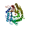
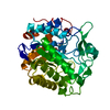
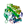
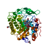
| Title | Crystal structure of Podospora anserina GH5 beta-(1,4)-mannanase | ||||||
 Components Components | GH5 ENDO-BETA-1,4-MANNANASE | ||||||
 Keywords Keywords | HYDROLASE / MANNANASE / GLYCOSYL HYDROLASE / CAZY | ||||||
| Function / homology |  Function and homology information Function and homology informationmannan catabolic process / mannan endo-1,4-beta-mannosidase / mannan endo-1,4-beta-mannosidase activity / extracellular region Similarity search - Function | ||||||
| Biological species |  PODOSPORA ANSERINA (fungus) PODOSPORA ANSERINA (fungus) | ||||||
| Method |  X-RAY DIFFRACTION / SYNCHROTRON / MOLECULAR REPLACEMENT / Resolution: 1.4 Å X-RAY DIFFRACTION / SYNCHROTRON / MOLECULAR REPLACEMENT / Resolution: 1.4 Å | ||||||
 Authors Authors | Couturier, M. / Roussel, A. / Rosengren, A. / Leone, P. / Stalbrand, H. / Berrin, J.G. | ||||||
 Citation Citation | Journal: J.Biol.Chem. / Year: 2013 Title: Structural and Biochemical Analyses of Glycoside Hydrolase Families 5 and 26 Beta-(1,4)-Mannanases from Podospora Anserina Reveal Differences Upon Manno-Oligosaccharides Catalysis. Authors: Couturier, M. / Roussel, A. / Rosengren, A. / Leone, P. / Stalbrand, H. / Berrin, J.G. | ||||||
| History |
|
- Structure visualization
| Structure viewer | Molecule: MolmilJmol/JSmol |
|---|
- Downloads & links
-Download
| PDBx/mmCIF format | 3ziz.cif.gz | 92.4 KB | Display | PDBx/mmCIF format |
|---|---|---|---|---|
| PDB format | pdb3ziz.ent.gz | 68.6 KB | Display | PDB format |
| PDBx/mmJSON format | 3ziz.json.gz | Tree view | PDBx/mmJSON format | |
| Others |  Other downloads Other downloads |
-Validation report
| Arichive directory | https://data.pdbj.org/pub/pdb/validation_reports/zi/3zizftp://data.pdbj.org/pub/pdb/validation_reports/zi/3ziz | HTTPS FTP |
|---|
-Related structure data
| Related structure data | 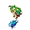 3zm8C  1qnoS C: citing same article ( S: Starting model for refinement |
|---|---|
| Similar structure data |


-Links
 PDBj
PDBj- Assembly
| Deposited unit | 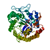
| ||||||||
|---|---|---|---|---|---|---|---|---|---|
| 1 |
| ||||||||
| Unit cell |
|
-Components
| #1: Protein | Mass: 42496.988 Da / Num. of mol.: 1 Source method: isolated from a genetically manipulated source Source: (gene. exp.) PODOSPORA ANSERINA (fungus) / Strain: S MAT / Plasmid: PPICZAA / Production host: PICHIA PASTORIS (fungus) / Strain (production host): X33References: UniProt: E2GHW1, UniProt: B2B3C0*PLUS, mannan endo-1,4-beta-mannosidase |
|---|---|
| #2: Chemical | ChemComp-GOL /   Mass: 92.094 Da / Num. of mol.: 1 / Source method: obtained synthetically / Formula: C3H8O3 Mass: 92.094 Da / Num. of mol.: 1 / Source method: obtained synthetically / Formula: C3H8O3 |
| #3: Chemical | ChemComp-TRS /   Mass: 122.143 Da / Num. of mol.: 1 / Source method: obtained synthetically / Formula: C4H12NO3 / Comment: pH buffer*YM Mass: 122.143 Da / Num. of mol.: 1 / Source method: obtained synthetically / Formula: C4H12NO3 / Comment: pH buffer*YM |
| #4: Water | ChemComp-HOH /  Mass: 18.015 Da / Num. of mol.: 434 / Source method: isolated from a natural source / Formula: H2O Mass: 18.015 Da / Num. of mol.: 434 / Source method: isolated from a natural source / Formula: H2O |
| Has protein modification | Y |
-Experimental details
-Experiment
| Experiment | Method: X-RAY DIFFRACTION / Number of used crystals: 1 |
|---|
- Sample preparation
| Crystal | Density Matthews: 2.32 Å3/Da / Density % sol: 47 % / Description: NONE |
|---|---|
| Crystal grow | pH: 8.5 Details: TRIS 0.1M PH 8.5, 0.2 M SODIUM ACETATE, 30% PEG 4000 |
-Data collection
| Diffraction | Mean temperature: 100 K |
|---|---|
| Diffraction source | Source: SYNCHROTRON / Site: ESRF  / Beamline: ID29 / Wavelength: 0.97914 / Beamline: ID29 / Wavelength: 0.97914 |
| Detector | Type: DECTRIS PILATUS 6M / Detector: PIXEL / Date: Oct 28, 2010 |
| Radiation | Protocol: SINGLE WAVELENGTH / Monochromatic (M) / Laue (L): M / Scattering type: x-ray |
| Radiation wavelength | Wavelength: 0.97914 Å / Relative weight: 1 |
| Reflection | Resolution: 1.4→30 Å / Num. obs: 62519 / % possible obs: 97.3 % / Observed criterion σ(I): 2 / Redundancy: 8.4 % / Biso Wilson estimate: 10.99 Å2 / Rmerge(I) obs: 0.09 / Net I/σ(I): 18.7 |
| Reflection shell | Resolution: 1.4→1.48 Å / Redundancy: 8.3 % / Rmerge(I) obs: 0.68 / Mean I/σ(I) obs: 3.4 / % possible all: 95.7 |
- Processing
| Software |
| ||||||||||||||||||||||||||||||||||||||||||||||||||||||||||||||||||||||||||||||||||||||||||||||||||||||||||||||||||
|---|---|---|---|---|---|---|---|---|---|---|---|---|---|---|---|---|---|---|---|---|---|---|---|---|---|---|---|---|---|---|---|---|---|---|---|---|---|---|---|---|---|---|---|---|---|---|---|---|---|---|---|---|---|---|---|---|---|---|---|---|---|---|---|---|---|---|---|---|---|---|---|---|---|---|---|---|---|---|---|---|---|---|---|---|---|---|---|---|---|---|---|---|---|---|---|---|---|---|---|---|---|---|---|---|---|---|---|---|---|---|---|---|---|---|---|
| Refinement | Method to determine structure: MOLECULAR REPLACEMENT Starting model: PDB ENTRY 1QNO Resolution: 1.4→28.95 Å / Cor.coef. Fo:Fc: 0.9657 / Cor.coef. Fo:Fc free: 0.9576 / SU R Cruickshank DPI: 0.054 / Cross valid method: THROUGHOUT / σ(F): 0 / SU R Blow DPI: 0.057 / SU Rfree Blow DPI: 0.057 / SU Rfree Cruickshank DPI: 0.055 Details: IDEAL-DIST CONTACT TERM CONTACT SETUP. RESIDUE TYPES WITHOUT CCP4 ATOM TYPE IN LIBRARY=. NUMBER OF ATOMS WITH PROPER CCP4 ATOM TYPE=3118. NUMBER WITH APPROX DEFAULT CCP4 ATOM TYPE=0. NUMBER ...Details: IDEAL-DIST CONTACT TERM CONTACT SETUP. RESIDUE TYPES WITHOUT CCP4 ATOM TYPE IN LIBRARY=. NUMBER OF ATOMS WITH PROPER CCP4 ATOM TYPE=3118. NUMBER WITH APPROX DEFAULT CCP4 ATOM TYPE=0. NUMBER TREATED BY BAD NON-BONDED CONTACTS=75.
| ||||||||||||||||||||||||||||||||||||||||||||||||||||||||||||||||||||||||||||||||||||||||||||||||||||||||||||||||||
| Displacement parameters | Biso mean: 13.05 Å2
| ||||||||||||||||||||||||||||||||||||||||||||||||||||||||||||||||||||||||||||||||||||||||||||||||||||||||||||||||||
| Refine analyze | Luzzati coordinate error obs: 0.126 Å | ||||||||||||||||||||||||||||||||||||||||||||||||||||||||||||||||||||||||||||||||||||||||||||||||||||||||||||||||||
| Refinement step | Cycle: LAST / Resolution: 1.4→28.95 Å
| ||||||||||||||||||||||||||||||||||||||||||||||||||||||||||||||||||||||||||||||||||||||||||||||||||||||||||||||||||
| Refine LS restraints |
| ||||||||||||||||||||||||||||||||||||||||||||||||||||||||||||||||||||||||||||||||||||||||||||||||||||||||||||||||||
| LS refinement shell | Resolution: 1.4→1.44 Å / Total num. of bins used: 20
|
