 Movie
Movie Controller

[English] 日本語
 Yorodumi
- PDB-5hos: Crystal structure of the endo-beta-1,4-glucanase Xac0029 from Xan... -
+ Open data

- Basic information
| Entry | Database: PDB / ID: 5hos | ||||||
|---|---|---|---|---|---|---|---|
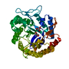
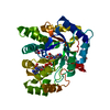
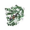
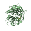
| Title | Crystal structure of the endo-beta-1,4-glucanase Xac0029 from Xanthomonas axonopodis pv. citri | ||||||
 Components Components | Cellulase | ||||||
 Keywords Keywords | HYDROLASE / endo-beta-1 / 4-glucanase | ||||||
| Function / homology |  Function and homology information Function and homology informationhydrolase activity, hydrolyzing O-glycosyl compounds / carbohydrate metabolic process Similarity search - Function | ||||||
| Biological species |  Xanthomonas axonopodis pv. citri (bacteria) Xanthomonas axonopodis pv. citri (bacteria) | ||||||
| Method |  X-RAY DIFFRACTION / SYNCHROTRON / MOLECULAR REPLACEMENT / Resolution: 1.6 Å X-RAY DIFFRACTION / SYNCHROTRON / MOLECULAR REPLACEMENT / Resolution: 1.6 Å | ||||||
 Authors Authors | Paiva, J.H. / Murakami, M.T. | ||||||
| Funding support |  Brazil, 1items Brazil, 1items
| ||||||
 Citation Citation | Journal: To Be Published Title: Crystal structure of the endo-beta-1,4-glucanase Xac0029 from Xanthomonas axonopodis pv. citri Authors: Paiva, J.H. / Murakami, M.T. | ||||||
| History |
|
- Structure visualization
| Structure viewer | Molecule: MolmilJmol/JSmol |
|---|
- Downloads & links
-Download
| PDBx/mmCIF format | 5hos.cif.gz | 144.2 KB | Display | PDBx/mmCIF format |
|---|---|---|---|---|
| PDB format | pdb5hos.ent.gz | 111.7 KB | Display | PDB format |
| PDBx/mmJSON format | 5hos.json.gz | Tree view | PDBx/mmJSON format | |
| Others |  Other downloads Other downloads |
-Validation report
| Arichive directory | https://data.pdbj.org/pub/pdb/validation_reports/ho/5hosftp://data.pdbj.org/pub/pdb/validation_reports/ho/5hos | HTTPS FTP |
|---|
-Related structure data
| Related structure data |  4w7uS S: Starting model for refinement |
|---|---|
| Similar structure data |


-Links
 PDBj
PDBj- Assembly
| Deposited unit | 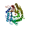
| ||||||||
|---|---|---|---|---|---|---|---|---|---|
| 1 |
| ||||||||
| Unit cell |
|
-Components
| #1: Protein | Mass: 37561.355 Da / Num. of mol.: 1 Source method: isolated from a genetically manipulated source Source: (gene. exp.) Xanthomonas axonopodis pv. citri (bacteria)Strain: 306 / Gene: egl, XAC0029 / Production host: |
|---|---|
| #2: Chemical | ChemComp-SO4 /   Mass: 96.063 Da / Num. of mol.: 1 / Source method: obtained synthetically / Formula: SO4 Mass: 96.063 Da / Num. of mol.: 1 / Source method: obtained synthetically / Formula: SO4 |
| #3: Water | ChemComp-HOH /  Mass: 18.015 Da / Num. of mol.: 198 / Source method: isolated from a natural source / Formula: H2O Mass: 18.015 Da / Num. of mol.: 198 / Source method: isolated from a natural source / Formula: H2O |
| Has protein modification | Y |
-Experimental details
-Experiment
| Experiment | Method: X-RAY DIFFRACTION / Number of used crystals: 1 |
|---|
- Sample preparation
| Crystal | Density Matthews: 2.38 Å3/Da / Density % sol: 48.26 % |
|---|---|
| Crystal grow | Temperature: 293 K / Method: vapor diffusion, sitting drop / Details: 25% PEG4000 0.2 M ammonium sulfate 15% glycerol |
-Data collection
| Diffraction | Mean temperature: 100 K | ||||||||||||||||||||||||||||||||||||||||||||||||||||||||||||
|---|---|---|---|---|---|---|---|---|---|---|---|---|---|---|---|---|---|---|---|---|---|---|---|---|---|---|---|---|---|---|---|---|---|---|---|---|---|---|---|---|---|---|---|---|---|---|---|---|---|---|---|---|---|---|---|---|---|---|---|---|---|
| Diffraction source | Source: SYNCHROTRON / Site: LNLS / Beamline: W01B-MX2 / Wavelength: 1.4587 Å | ||||||||||||||||||||||||||||||||||||||||||||||||||||||||||||
| Detector | Type: DECTRIS PILATUS 2M / Detector: PIXEL / Date: Nov 16, 2015 | ||||||||||||||||||||||||||||||||||||||||||||||||||||||||||||
| Radiation | Protocol: SINGLE WAVELENGTH / Monochromatic (M) / Laue (L): M / Scattering type: x-ray | ||||||||||||||||||||||||||||||||||||||||||||||||||||||||||||
| Radiation wavelength | Wavelength: 1.4587 Å / Relative weight: 1 | ||||||||||||||||||||||||||||||||||||||||||||||||||||||||||||
| Reflection | Resolution: 1.6→42.8 Å / Num. obs: 47130 / % possible obs: 99.8 % / Observed criterion σ(I): -3 / Redundancy: 7.06 % / Biso Wilson estimate: 29.143 Å2 / Rmerge(I) obs: 0.067 / Net I/σ(I): 15.14 | ||||||||||||||||||||||||||||||||||||||||||||||||||||||||||||
| Reflection shell |
|
- Processing
| Software |
| ||||||||||||||||||||||||||||||||||||||||||||||||||||||||||||||||||||||||||||||||||||||||||
|---|---|---|---|---|---|---|---|---|---|---|---|---|---|---|---|---|---|---|---|---|---|---|---|---|---|---|---|---|---|---|---|---|---|---|---|---|---|---|---|---|---|---|---|---|---|---|---|---|---|---|---|---|---|---|---|---|---|---|---|---|---|---|---|---|---|---|---|---|---|---|---|---|---|---|---|---|---|---|---|---|---|---|---|---|---|---|---|---|---|---|---|
| Refinement | Method to determine structure: MOLECULAR REPLACEMENT Starting model: 4W7U Resolution: 1.6→42.8 Å / Cor.coef. Fo:Fc: 0.97 / Cor.coef. Fo:Fc free: 0.961 / SU B: 5.385 / SU ML: 0.076 / Cross valid method: THROUGHOUT / σ(F): 0 / ESU R: 0.098 / ESU R Free: 0.081 Details: HYDROGENS HAVE BEEN ADDED IN THE RIDING POSITIONS U VALUES : REFINED INDIVIDUALLY
| ||||||||||||||||||||||||||||||||||||||||||||||||||||||||||||||||||||||||||||||||||||||||||
| Solvent computation | Ion probe radii: 0.8 Å / Shrinkage radii: 0.8 Å / VDW probe radii: 1.2 Å | ||||||||||||||||||||||||||||||||||||||||||||||||||||||||||||||||||||||||||||||||||||||||||
| Displacement parameters | Biso max: 64.92 Å2 / Biso mean: 23.898 Å2 / Biso min: 12.57 Å2
| ||||||||||||||||||||||||||||||||||||||||||||||||||||||||||||||||||||||||||||||||||||||||||
| Refinement step | Cycle: final / Resolution: 1.6→42.8 Å
| ||||||||||||||||||||||||||||||||||||||||||||||||||||||||||||||||||||||||||||||||||||||||||
| Refine LS restraints |
| ||||||||||||||||||||||||||||||||||||||||||||||||||||||||||||||||||||||||||||||||||||||||||
| LS refinement shell | Resolution: 1.604→1.646 Å / Total num. of bins used: 20
|
