 Movie
Movie Controller


+ Open data

- Basic information
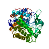
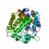
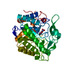
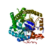
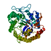
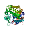
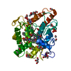
| Entry | Database: PDB / ID: 1h51 | ||||||
|---|---|---|---|---|---|---|---|
| Title | Oxidised Pentaerythritol Tetranitrate Reductase (SCN complex) | ||||||
 Components Components | PENTAERYTHRITOL TETRANITRATE REDUCTASE | ||||||
 Keywords Keywords | OXIDOREDUCTASE / FLAVOENZYME / EXPLOSIVE DEGRADATION / STEROID BINDING | ||||||
| Function / homology |  Function and homology information Function and homology informationoxidoreductase activity, acting on the CH-CH group of donors, NAD or NADP as acceptor / FMN binding / cytosol Similarity search - Function | ||||||
| Biological species |  ENTEROBACTER CLOACAE (bacteria) ENTEROBACTER CLOACAE (bacteria) | ||||||
| Method |  X-RAY DIFFRACTION / MOLECULAR REPLACEMENT / Resolution: 1.6 Å X-RAY DIFFRACTION / MOLECULAR REPLACEMENT / Resolution: 1.6 Å | ||||||
 Authors Authors | Barna, T. / Moody, P.C.E. | ||||||
 Citation Citation | Journal: J.Mol.Biol. / Year: 2001 Title: Crystal Structure of Pentaerythritol Tetranitrate Reductase: "Flipped" Binding Geometries for Steroid Substrates in Different Redox States of the Enzyme Authors: Barna, T.M. / Khan, H. / Bruce, N.C. / Barsukov, I. / Scrutton, N.S. / Moody, P.C.E. #1: Journal: Acta Crystallogr.,Sect.D / Year: 1998 Title: Crystallisation and Preliminary Diffraction Studies of Pentaerythritol Reductase from Enterobacter Cloacae Pb2 Authors: Moody, P.C.E. / Shikotra, N. / French, C.E. / Bruce, N.C. / Scrutton, N.S. | ||||||
| History |
| ||||||
| Remark 700 | SHEET DETERMINATION METHOD: DSSP THE SHEETS PRESENTED AS "A2" IN EACH CHAIN ON SHEET RECORDS BELOW ... SHEET DETERMINATION METHOD: DSSP THE SHEETS PRESENTED AS "A2" IN EACH CHAIN ON SHEET RECORDS BELOW IS ACTUALLY AN 8-STRANDED BARREL THIS IS REPRESENTED BY A 9-STRANDED SHEET IN WHICH THE FIRST AND LAST STRANDS ARE IDENTICAL. |
- Structure visualization
| Structure viewer | Molecule: MolmilJmol/JSmol |
|---|
- Downloads & links
-Download
| PDBx/mmCIF format | 1h51.cif.gz | 93.1 KB | Display | PDBx/mmCIF format |
|---|---|---|---|---|
| PDB format | pdb1h51.ent.gz | 69 KB | Display | PDB format |
| PDBx/mmJSON format | 1h51.json.gz | Tree view | PDBx/mmJSON format | |
| Others |  Other downloads Other downloads |
-Validation report
| Arichive directory | https://data.pdbj.org/pub/pdb/validation_reports/h5/1h51ftp://data.pdbj.org/pub/pdb/validation_reports/h5/1h51 | HTTPS FTP |
|---|
-Related structure data
| Related structure data | 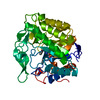 1h50C  1h60C  1h61C  1h62C  1h63C  1oyaS C: citing same article ( S: Starting model for refinement |
|---|---|
| Similar structure data |


-Links
 PDBj
PDBj
- Assembly
| Deposited unit | 
| ||||||||
|---|---|---|---|---|---|---|---|---|---|
| 1 |
| ||||||||
| Unit cell |
|
-Components
| #1: Protein | Mass: 39404.000 Da / Num. of mol.: 1 Source method: isolated from a genetically manipulated source Details: THIOCYANATE IS BOUND IN THE ACTIVE SITE / Source: (gene. exp.) ENTEROBACTER CLOACAE (bacteria) / Strain: PB2 / Description: NCBI U68759. RECOMBINANT / Plasmid: PONR1 / Production host: |
|---|---|
| #2: Chemical | ChemComp-FMN /   Mass: 456.344 Da / Num. of mol.: 1 / Source method: obtained synthetically / Formula: C17H21N4O9P Mass: 456.344 Da / Num. of mol.: 1 / Source method: obtained synthetically / Formula: C17H21N4O9P |
| #3: Chemical | ChemComp-SCN /   Mass: 58.082 Da / Num. of mol.: 1 / Source method: obtained synthetically / Formula: CNS Mass: 58.082 Da / Num. of mol.: 1 / Source method: obtained synthetically / Formula: CNS |
| #4: Water | ChemComp-HOH /  Mass: 18.015 Da / Num. of mol.: 476 / Source method: isolated from a natural source / Formula: H2O Mass: 18.015 Da / Num. of mol.: 476 / Source method: isolated from a natural source / Formula: H2O |
-Experimental details
-Experiment
| Experiment | Method: X-RAY DIFFRACTION / Number of used crystals: 1 |
|---|
- Sample preparation
| Crystal | Density Matthews: 2.3 Å3/Da / Density % sol: 38 % |
|---|---|
| Crystal grow | pH: 6.2 Details: 25% PEG 3000, 0.05M SODIUM THIOCYANATE, 0.1M SODIUM CACODYLATE, 17% ISOPROPANOL PH 6.2 |
-Data collection
| Diffraction | Mean temperature: 100 K |
|---|---|
| Diffraction source | Source: ROTATING ANODE / Type: RIGAKU RU200 / Wavelength: 1.5418 |
| Detector | Type: RIGAKU RAXIS IV IMAGE PLATE / Detector: IMAGE PLATE / Details: MIRRORS |
| Radiation | Monochromator: NI FILTER / Protocol: SINGLE WAVELENGTH / Monochromatic (M) / Laue (L): M / Scattering type: x-ray |
| Radiation wavelength | Wavelength: 1.5418 Å / Relative weight: 1 |
| Reflection | Resolution: 1.6→50 Å / Num. obs: 51016 / % possible obs: 93.7 % / Redundancy: 10.2 % / Rmerge(I) obs: 0.027 / Net I/σ(I): 24.1 |
- Processing
| Software |
| ||||||||||||||||||||||||||||||||||||||||||||||||||||||||||||
|---|---|---|---|---|---|---|---|---|---|---|---|---|---|---|---|---|---|---|---|---|---|---|---|---|---|---|---|---|---|---|---|---|---|---|---|---|---|---|---|---|---|---|---|---|---|---|---|---|---|---|---|---|---|---|---|---|---|---|---|---|---|
| Refinement | Method to determine structure: MOLECULAR REPLACEMENT Starting model: PDB ENTRY 1OYA Resolution: 1.6→50 Å / Cross valid method: THROUGHOUT / σ(F): 0
| ||||||||||||||||||||||||||||||||||||||||||||||||||||||||||||
| Refinement step | Cycle: LAST / Resolution: 1.6→50 Å
| ||||||||||||||||||||||||||||||||||||||||||||||||||||||||||||
| Refine LS restraints |
|
