 Movie
Movie Controller

[English] 日本語
 Yorodumi
- PDB-1vyp: Structure of pentaerythritol tetranitrate reductase W102F mutant ... -
+ Open data

- Basic information
| Entry | Database: PDB / ID: 1vyp | ||||||
|---|---|---|---|---|---|---|---|
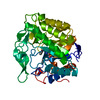
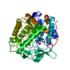
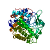
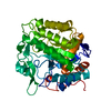
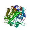
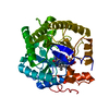
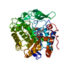
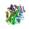
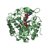
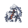
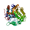
| Title | Structure of pentaerythritol tetranitrate reductase W102F mutant and complexed with picric acid | ||||||
 Components Components | PENTAERYTHRITOL TETRANITRATE REDUCTASE | ||||||
 Keywords Keywords | OXIDOREDUCTASE / FLAVOENZYME / EXPLOSIVE DEGRADATION / STEROID BINDING | ||||||
| Function / homology |  Function and homology information Function and homology informationoxidoreductase activity, acting on the CH-CH group of donors, NAD or NADP as acceptor / FMN binding / cytosol Similarity search - Function | ||||||
| Biological species |  ENTEROBACTER CLOACAE (bacteria) ENTEROBACTER CLOACAE (bacteria) | ||||||
| Method |  X-RAY DIFFRACTION / SYNCHROTRON / MOLECULAR REPLACEMENT / Resolution: 1.27 Å X-RAY DIFFRACTION / SYNCHROTRON / MOLECULAR REPLACEMENT / Resolution: 1.27 Å | ||||||
 Authors Authors | Barna, T. / Moody, P.C.E. | ||||||
 Citation Citation | Journal: J.Biol.Chem. / Year: 2004 Title: Atomic Resolution Structures and Solution Behavior of Enzyme-Substrate Complexes of Enterobacter Cloacae Pb2 Pentaerythritol Tetranitrate Reductase: Multiple Conformational States and ...Title: Atomic Resolution Structures and Solution Behavior of Enzyme-Substrate Complexes of Enterobacter Cloacae Pb2 Pentaerythritol Tetranitrate Reductase: Multiple Conformational States and Implications for the Mechanism of Nitroaromatic Explosive Degradation Authors: Khan, H. / Barna, T. / Harris, R. / Bruce, N. / Barsukov, I. / Munro, A. / Moody, P.C.E. / Scrutton, N. #1: Journal: J.Mol.Biol. / Year: 2001Title: Crystal Structure of Pentaerythritol Tetranitrate Reductase: "Flipped" Binding Geometries for Steroid Substrates in Different Redox States of the Enzyme Authors: Barna, T.M. / Khan, H. / Bruce, N.C. / Barsukov, I. / Scrutton, N.S. / Moody, P.C. #2: Journal: Acta Crystallogr.,Sect.D / Year: 1998 Title: Crystallization and Preliminary Diffraction Studies of Pentaerythritol Tetranitrate Reductase from Enterobacter Cloacae Pb2. Authors: Moody, P.C.E. / Shikotra, N. / French, C.E. / Bruce, N.C. / Scrutton, N.S. | ||||||
| History |
| ||||||
| Remark 700 | SHEET DETERMINATION METHOD: DSSP THE SHEETS PRESENTED AS "XB" IN EACH CHAIN ON SHEET RECORDS BELOW ... SHEET DETERMINATION METHOD: DSSP THE SHEETS PRESENTED AS "XB" IN EACH CHAIN ON SHEET RECORDS BELOW IS ACTUALLY AN 8-STRANDED BARREL THIS IS REPRESENTED BY A 9-STRANDED SHEET IN WHICH THE FIRST AND LAST STRANDS ARE IDENTICAL. |
- Structure visualization
| Structure viewer | Molecule: MolmilJmol/JSmol |
|---|
- Downloads & links
-Download
| PDBx/mmCIF format | 1vyp.cif.gz | 178.7 KB | Display | PDBx/mmCIF format |
|---|---|---|---|---|
| PDB format | pdb1vyp.ent.gz | 139.4 KB | Display | PDB format |
| PDBx/mmJSON format | 1vyp.json.gz | Tree view | PDBx/mmJSON format | |
| Others |  Other downloads Other downloads |
-Validation report
| Arichive directory | https://data.pdbj.org/pub/pdb/validation_reports/vy/1vypftp://data.pdbj.org/pub/pdb/validation_reports/vy/1vyp | HTTPS FTP |
|---|
-Related structure data
| Related structure data |  1vyrC  1vysC  1gvsS S: Starting model for refinement C: citing same article ( |
|---|---|
| Similar structure data |


-Links
 PDBj
PDBj- Assembly
| Deposited unit | 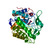
| ||||||||
|---|---|---|---|---|---|---|---|---|---|
| 1 |
| ||||||||
| Unit cell |
|
-Components
| #1: Protein | Mass: 39364.965 Da / Num. of mol.: 1 / Mutation: YES Source method: isolated from a genetically manipulated source Details: 2,4,6 TRINITROPHENOL IS BOUND IN THE ACTIVE SITE / Source: (gene. exp.) ENTEROBACTER CLOACAE (bacteria) / Description: NCBI U68759. RECOMBINANT / Plasmid: PONR1 / Production host: |
|---|---|
| #2: Chemical | ChemComp-FMN /   Mass: 456.344 Da / Num. of mol.: 1 / Source method: obtained synthetically / Formula: C17H21N4O9P Mass: 456.344 Da / Num. of mol.: 1 / Source method: obtained synthetically / Formula: C17H21N4O9P |
| #3: Chemical | ChemComp-TNF /   Mass: 229.104 Da / Num. of mol.: 1 / Source method: obtained synthetically / Formula: C6H3N3O7 Mass: 229.104 Da / Num. of mol.: 1 / Source method: obtained synthetically / Formula: C6H3N3O7 |
| #4: Water | ChemComp-HOH /  Mass: 18.015 Da / Num. of mol.: 705 / Source method: isolated from a natural source / Formula: H2O Mass: 18.015 Da / Num. of mol.: 705 / Source method: isolated from a natural source / Formula: H2O |
| Compound details | ENGINEERED |
-Experimental details
-Experiment
| Experiment | Method: X-RAY DIFFRACTION / Number of used crystals: 1 |
|---|
- Sample preparation
| Crystal | Density Matthews: 2.3 Å3/Da / Density % sol: 46.63 % |
|---|---|
| Crystal grow | pH: 6.2 / Details: pH 6.20 |
-Data collection
| Diffraction | Mean temperature: 100 K |
|---|---|
| Diffraction source | Source: SYNCHROTRON / Site: SRS  / Beamline: PX9.6 / Wavelength: 0.87 / Beamline: PX9.6 / Wavelength: 0.87 |
| Detector | Type: ADSC CCD / Detector: CCD / Date: Jan 12, 1999 |
| Radiation | Protocol: SINGLE WAVELENGTH / Monochromatic (M) / Laue (L): M / Scattering type: x-ray |
| Radiation wavelength | Wavelength: 0.87 Å / Relative weight: 1 |
| Reflection | Resolution: 1.27→50 Å / Num. obs: 29340 / % possible obs: 93.8 % / Redundancy: 3.64 % / Rmerge(I) obs: 0.055 / Net I/σ(I): 17.1 |
| Reflection shell | Resolution: 1.27→1.32 Å / Rmerge(I) obs: 0.26 / Mean I/σ(I) obs: 2.5 / % possible all: 90.4 |
- Processing
| Software |
| ||||||||||||||||||||
|---|---|---|---|---|---|---|---|---|---|---|---|---|---|---|---|---|---|---|---|---|---|
| Refinement | Method to determine structure: MOLECULAR REPLACEMENT Starting model: PDB ENTRY 1GVS Resolution: 1.27→37.8 Å / SU B: 0.549 / SU ML: 0.024 / Cross valid method: THROUGHOUT / σ(F): 0 / ESU R: 0.046 / ESU R Free: 0.042
| ||||||||||||||||||||
| Displacement parameters | Biso mean: 9.56 Å2
| ||||||||||||||||||||
| Refinement step | Cycle: LAST / Resolution: 1.27→37.8 Å
|
