 Movie
Movie Controller

[English] 日本語
 Yorodumi
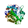
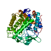
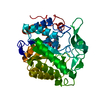
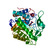
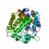
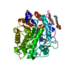
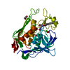
- PDB-3p82: H184N mutant of pentaerythritol tetranitrate reductase containing... -
+ Open data

- Basic information
| Entry | Database: PDB / ID: 3p82 | ||||||
|---|---|---|---|---|---|---|---|
| Title | H184N mutant of pentaerythritol tetranitrate reductase containing bound acetate ion | ||||||
 Components Components | Pentaerythritol tetranitrate reductase | ||||||
 Keywords Keywords | OXIDOREDUCTASE / old yellow enzyme family / alpha / beta barrel | ||||||
| Function / homology |  Function and homology information Function and homology informationoxidoreductase activity, acting on the CH-CH group of donors, NAD or NADP as acceptor / FMN binding / cytosol Similarity search - Function | ||||||
| Biological species |  Enterobacter cloacae (bacteria) Enterobacter cloacae (bacteria) | ||||||
| Method |  X-RAY DIFFRACTION / MOLECULAR REPLACEMENT / molecular replacement / Resolution: 2.2 Å X-RAY DIFFRACTION / MOLECULAR REPLACEMENT / molecular replacement / Resolution: 2.2 Å | ||||||
 Authors Authors | Toogood, H.S. / Scrutton, N.S. | ||||||
 Citation Citation | Journal: Chembiochem / Year: 2011 Title: A Site-Saturated Mutagenesis Study of Pentaerythritol Tetranitrate Reductase Reveals that Residues 181 and 184 Influence Ligand Binding, Stereochemistry and Reactivity. Authors: Toogood, H.S. / Fryszkowska, A. / Hulley, M. / Sakuma, M. / Mansell, D. / Stephens, G.M. / Gardiner, J.M. / Scrutton, N.S. | ||||||
| History |
|
- Structure visualization
| Structure viewer | Molecule: MolmilJmol/JSmol |
|---|
- Downloads & links
-Download
| PDBx/mmCIF format | 3p82.cif.gz | 98.2 KB | Display | PDBx/mmCIF format |
|---|---|---|---|---|
| PDB format | pdb3p82.ent.gz | 71.7 KB | Display | PDB format |
| PDBx/mmJSON format | 3p82.json.gz | Tree view | PDBx/mmJSON format | |
| Others |  Other downloads Other downloads |
-Validation report
| Arichive directory | https://data.pdbj.org/pub/pdb/validation_reports/p8/3p82ftp://data.pdbj.org/pub/pdb/validation_reports/p8/3p82 | HTTPS FTP |
|---|
-Related structure data
| Related structure data | 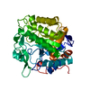 3p74C 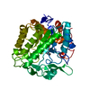 3p7yC 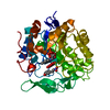 3p80C 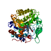 3p81C 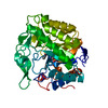 1h50S C: citing same article ( S: Starting model for refinement |
|---|---|
| Similar structure data |


-Links
 PDBj
PDBj- Assembly
| Deposited unit | 
| ||||||||
|---|---|---|---|---|---|---|---|---|---|
| 1 |
| ||||||||
| Unit cell |
|
-Components
| #1: Protein | Mass: 39511.152 Da / Num. of mol.: 1 / Mutation: H184N Source method: isolated from a genetically manipulated source Source: (gene. exp.) Enterobacter cloacae (bacteria) / Strain: PB2 / Gene: onr, PETNR / Plasmid: pBluescript II-KS(+) / Production host: |
|---|---|
| #2: Chemical | ChemComp-FMN /   Mass: 456.344 Da / Num. of mol.: 1 / Source method: obtained synthetically / Formula: C17H21N4O9P Mass: 456.344 Da / Num. of mol.: 1 / Source method: obtained synthetically / Formula: C17H21N4O9P |
| #3: Chemical | ChemComp-ACT /   Mass: 59.044 Da / Num. of mol.: 1 / Source method: obtained synthetically / Formula: C2H3O2 Mass: 59.044 Da / Num. of mol.: 1 / Source method: obtained synthetically / Formula: C2H3O2 |
| #4: Water | ChemComp-HOH /  Mass: 18.015 Da / Num. of mol.: 524 / Source method: isolated from a natural source / Formula: H2O Mass: 18.015 Da / Num. of mol.: 524 / Source method: isolated from a natural source / Formula: H2O |
-Experimental details
-Experiment
| Experiment | Method: X-RAY DIFFRACTION / Number of used crystals: 1 |
|---|
- Sample preparation
| Crystal | Density Matthews: 2.19 Å3/Da / Density % sol: 43.71 % |
|---|---|
| Crystal grow | Temperature: 293 K / Method: vapor diffusion, sitting drop / pH: 6.2 Details: 100mM sodium cacodylate, 100 mM sodium acetate, 16-18% isopropanol, pH 6.2, VAPOR DIFFUSION, SITTING DROP, temperature 293K |
-Data collection
| Diffraction | Mean temperature: 100 K |
|---|---|
| Diffraction source | Source: ROTATING ANODE / Type: BRUKER AXS MICROSTAR / Wavelength: 1.542 Å |
| Detector | Type: X8 Proteum / Detector: CCD / Date: Mar 11, 2009 |
| Radiation | Monochromator: Montel 200 Mirror optics / Protocol: SINGLE WAVELENGTH / Monochromatic (M) / Laue (L): M / Scattering type: x-ray |
| Radiation wavelength | Wavelength: 1.542 Å / Relative weight: 1 |
| Reflection | Resolution: 2.2→26.4 Å / Num. all: 28870 / Num. obs: 28870 / % possible obs: 99.9 % / Redundancy: 10.9 % / Rmerge(I) obs: 0.092 / Rsym value: 0.092 / Net I/σ(I): 17.2 |
| Reflection shell | Resolution: 2.252→2.393 Å / Redundancy: 11.33 % / Mean I/σ(I) obs: 3.4 / Rsym value: 0.113 / % possible all: 99.89 |
-Phasing
| Phasing | Method: molecular replacement |
|---|
- Processing
| Software |
| ||||||||||||||||||||||||||||||||||||||||||||||||||||||||||||||||||||||||||||||||||||||||||
|---|---|---|---|---|---|---|---|---|---|---|---|---|---|---|---|---|---|---|---|---|---|---|---|---|---|---|---|---|---|---|---|---|---|---|---|---|---|---|---|---|---|---|---|---|---|---|---|---|---|---|---|---|---|---|---|---|---|---|---|---|---|---|---|---|---|---|---|---|---|---|---|---|---|---|---|---|---|---|---|---|---|---|---|---|---|---|---|---|---|---|---|
| Refinement | Method to determine structure: MOLECULAR REPLACEMENT Starting model: 1H50 Resolution: 2.2→26.4 Å / Cor.coef. Fo:Fc: 0.965 / Cor.coef. Fo:Fc free: 0.915 / WRfactor Rfree: 0.2103 / WRfactor Rwork: 0.1402 / Occupancy max: 1 / Occupancy min: 0.5 / FOM work R set: 0.8425 / SU B: 5.294 / SU ML: 0.136 / SU R Cruickshank DPI: 0.3013 / SU Rfree: 0.2189 / Cross valid method: THROUGHOUT / σ(F): 0 / ESU R Free: 0.219 / Stereochemistry target values: MAXIMUM LIKELIHOOD / Details: HYDROGENS HAVE BEEN ADDED IN THE RIDING POSITIONS
| ||||||||||||||||||||||||||||||||||||||||||||||||||||||||||||||||||||||||||||||||||||||||||
| Solvent computation | Ion probe radii: 0.8 Å / Shrinkage radii: 0.8 Å / VDW probe radii: 1.2 Å / Solvent model: MASK | ||||||||||||||||||||||||||||||||||||||||||||||||||||||||||||||||||||||||||||||||||||||||||
| Displacement parameters | Biso max: 61.4 Å2 / Biso mean: 19.1172 Å2 / Biso min: 2.14 Å2
| ||||||||||||||||||||||||||||||||||||||||||||||||||||||||||||||||||||||||||||||||||||||||||
| Refinement step | Cycle: LAST / Resolution: 2.2→26.4 Å
| ||||||||||||||||||||||||||||||||||||||||||||||||||||||||||||||||||||||||||||||||||||||||||
| Refine LS restraints |
| ||||||||||||||||||||||||||||||||||||||||||||||||||||||||||||||||||||||||||||||||||||||||||
| LS refinement shell | Resolution: 2.2→2.257 Å / Total num. of bins used: 20
|
