 Movie
Movie Controller

[English] 日本語
 Yorodumi
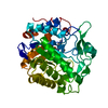
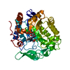
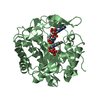
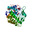
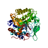
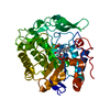
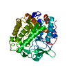
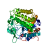
- PDB-6gi9: Crystal structure of pentaerythritol tetranitrate reductase (PETN... -
+ Open data

- Basic information
| Entry | Database: PDB / ID: 6gi9 | ||||||||||||
|---|---|---|---|---|---|---|---|---|---|---|---|---|---|
| Title | Crystal structure of pentaerythritol tetranitrate reductase (PETNR) mutant I107L | ||||||||||||
 Components Components | Pentaerythritol tetranitrate reductase | ||||||||||||
 Keywords Keywords | FLAVOPROTEIN / Mutant I107L | ||||||||||||
| Function / homology |  Function and homology information Function and homology informationoxidoreductase activity, acting on the CH-CH group of donors, NAD or NADP as acceptor / FMN binding / cytosol Similarity search - Function | ||||||||||||
| Biological species |  Enterobacter cloacae (bacteria) Enterobacter cloacae (bacteria) | ||||||||||||
| Method |  X-RAY DIFFRACTION / SYNCHROTRON / MOLECULAR REPLACEMENT / Resolution: 1.45 Å X-RAY DIFFRACTION / SYNCHROTRON / MOLECULAR REPLACEMENT / Resolution: 1.45 Å | ||||||||||||
 Authors Authors | Levy, C.W. | ||||||||||||
| Funding support |  United Kingdom, 3items United Kingdom, 3items
| ||||||||||||
 Citation Citation | Journal: Acs Catalysis / Year: 2018 Title: Nonequivalence of Second Sphere "Noncatalytic" Residues in Pentaerythritol Tetranitrate Reductase in Relation to Local Dynamics Linked to H-Transfer in Reactions with NADH and NADPH Coenzymes. Authors: Iorgu, A.I. / Baxter, N.J. / Cliff, M.J. / Levy, C. / Waltho, J.P. / Hay, S. / Scrutton, N.S. | ||||||||||||
| History |
|
- Structure visualization
| Structure viewer | Molecule: MolmilJmol/JSmol |
|---|
- Downloads & links
-Download
| PDBx/mmCIF format | 6gi9.cif.gz | 275.3 KB | Display | PDBx/mmCIF format |
|---|---|---|---|---|
| PDB format | pdb6gi9.ent.gz | 182.8 KB | Display | PDB format |
| PDBx/mmJSON format | 6gi9.json.gz | Tree view | PDBx/mmJSON format | |
| Others |  Other downloads Other downloads |
-Validation report
| Arichive directory | https://data.pdbj.org/pub/pdb/validation_reports/gi/6gi9ftp://data.pdbj.org/pub/pdb/validation_reports/gi/6gi9 | HTTPS FTP |
|---|
-Related structure data
| Related structure data |  6gi7C  6gi8C  6giaC  2abaS S: Starting model for refinement C: citing same article ( |
|---|---|
| Similar structure data |


-Links
 PDBj
PDBj- Assembly
| Deposited unit | 
| ||||||||||||
|---|---|---|---|---|---|---|---|---|---|---|---|---|---|
| 1 |
| ||||||||||||
| Unit cell |
|
-Components
| #1: Protein | Mass: 40640.359 Da / Num. of mol.: 1 Source method: isolated from a genetically manipulated source Source: (gene. exp.) Enterobacter cloacae (bacteria) / Gene: onr / Production host: |
|---|---|
| #2: Chemical | ChemComp-FMN /   Mass: 456.344 Da / Num. of mol.: 1 / Source method: obtained synthetically / Formula: C17H21N4O9P Mass: 456.344 Da / Num. of mol.: 1 / Source method: obtained synthetically / Formula: C17H21N4O9P |
| #3: Chemical | ChemComp-ACT /   Mass: 59.044 Da / Num. of mol.: 1 / Source method: isolated from a natural source / Formula: C2H3O2 Mass: 59.044 Da / Num. of mol.: 1 / Source method: isolated from a natural source / Formula: C2H3O2 |
| #4: Water | ChemComp-HOH /  Mass: 18.015 Da / Num. of mol.: 508 / Source method: isolated from a natural source / Formula: H2O Mass: 18.015 Da / Num. of mol.: 508 / Source method: isolated from a natural source / Formula: H2O |
-Experimental details
-Experiment
| Experiment | Method: X-RAY DIFFRACTION / Number of used crystals: 1 |
|---|
- Sample preparation
| Crystal | Density Matthews: 2.25 Å3/Da / Density % sol: 45.44 % |
|---|---|
| Crystal grow | Temperature: 294 K / Method: vapor diffusion, sitting drop / pH: 8 Details: 25 % (w/v) PEG 3000, 17 % (v/v) isopropanol, 0.1 M trisodium citrate, 0.1 M cacodylic acid (pH 6.5) |
-Data collection
| Diffraction | Mean temperature: 100 K |
|---|---|
| Diffraction source | Source: SYNCHROTRON / Site: Diamond / Beamline: I24 / Wavelength: 0.969 Å |
| Detector | Type: DECTRIS PILATUS3 6M / Detector: PIXEL / Date: Oct 18, 2013 |
| Radiation | Protocol: SINGLE WAVELENGTH / Monochromatic (M) / Laue (L): M / Scattering type: x-ray |
| Radiation wavelength | Wavelength: 0.969 Å / Relative weight: 1 |
| Reflection | Resolution: 1.45→37.54 Å / Num. obs: 63458 / % possible obs: 98.68 % / Redundancy: 3.6 % / Biso Wilson estimate: 13.03 Å2 / CC1/2: 0.998 / Rmerge(I) obs: 0.0633 / Rpim(I) all: 0.0378 / Rrim(I) all: 0.074 / Net I/σ(I): 13.19 |
| Reflection shell | Resolution: 1.45→1.502 Å / Redundancy: 3.5 % / Rmerge(I) obs: 0.604 / Mean I/σ(I) obs: 2.16 / Num. unique obs: 6292 / CC1/2: 0.728 / Rpim(I) all: 0.37 / Rrim(I) all: 0.713 / % possible all: 99.31 |
- Processing
| Software |
| ||||||||||||||||||||||||||||||||||||||||||||||||||||||||||||||||||||||||||||||||||||||||||||||||||||||||||||||||||||||||||||||||||||||||||||||||||||||||||||||||||||||||
|---|---|---|---|---|---|---|---|---|---|---|---|---|---|---|---|---|---|---|---|---|---|---|---|---|---|---|---|---|---|---|---|---|---|---|---|---|---|---|---|---|---|---|---|---|---|---|---|---|---|---|---|---|---|---|---|---|---|---|---|---|---|---|---|---|---|---|---|---|---|---|---|---|---|---|---|---|---|---|---|---|---|---|---|---|---|---|---|---|---|---|---|---|---|---|---|---|---|---|---|---|---|---|---|---|---|---|---|---|---|---|---|---|---|---|---|---|---|---|---|---|---|---|---|---|---|---|---|---|---|---|---|---|---|---|---|---|---|---|---|---|---|---|---|---|---|---|---|---|---|---|---|---|---|---|---|---|---|---|---|---|---|---|---|---|---|---|---|---|---|
| Refinement | Method to determine structure: MOLECULAR REPLACEMENT Starting model: 2ABA Resolution: 1.45→37.54 Å / SU ML: 0.1354 / Cross valid method: FREE R-VALUE / σ(F): 1.34 / Phase error: 16.1287
| ||||||||||||||||||||||||||||||||||||||||||||||||||||||||||||||||||||||||||||||||||||||||||||||||||||||||||||||||||||||||||||||||||||||||||||||||||||||||||||||||||||||||
| Solvent computation | Shrinkage radii: 0.9 Å / VDW probe radii: 1.11 Å | ||||||||||||||||||||||||||||||||||||||||||||||||||||||||||||||||||||||||||||||||||||||||||||||||||||||||||||||||||||||||||||||||||||||||||||||||||||||||||||||||||||||||
| Displacement parameters | Biso mean: 16.75 Å2 | ||||||||||||||||||||||||||||||||||||||||||||||||||||||||||||||||||||||||||||||||||||||||||||||||||||||||||||||||||||||||||||||||||||||||||||||||||||||||||||||||||||||||
| Refinement step | Cycle: LAST / Resolution: 1.45→37.54 Å
| ||||||||||||||||||||||||||||||||||||||||||||||||||||||||||||||||||||||||||||||||||||||||||||||||||||||||||||||||||||||||||||||||||||||||||||||||||||||||||||||||||||||||
| Refine LS restraints |
| ||||||||||||||||||||||||||||||||||||||||||||||||||||||||||||||||||||||||||||||||||||||||||||||||||||||||||||||||||||||||||||||||||||||||||||||||||||||||||||||||||||||||
| LS refinement shell |
|
