 Movie
Movie Controller

[English] 日本語
 Yorodumi
- PDB-5fml: Crystal structure of the endonuclease from the PA subunit of infl... -
+ Open data

- Basic information
| Entry | Database: PDB / ID: 5fml | ||||||
|---|---|---|---|---|---|---|---|
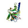
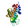
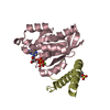
| Title | Crystal structure of the endonuclease from the PA subunit of influenza B virus bound to the PB2 subunit NLS peptide | ||||||
 Components Components |
| ||||||
 Keywords Keywords | VIRAL PROTEIN / ENDONUCLEASE | ||||||
| Function / homology |  Function and homology information Function and homology informationcap snatching / symbiont-mediated suppression of host mRNA transcription via inhibition of RNA polymerase II activity / host cell mitochondrion / 7-methylguanosine mRNA capping / virion component / endonuclease activity / Hydrolases; Acting on ester bonds / host cell cytoplasm / symbiont-mediated suppression of host gene expression / viral translational frameshifting ...cap snatching / symbiont-mediated suppression of host mRNA transcription via inhibition of RNA polymerase II activity / host cell mitochondrion / 7-methylguanosine mRNA capping / virion component / endonuclease activity / Hydrolases; Acting on ester bonds / host cell cytoplasm / symbiont-mediated suppression of host gene expression / viral translational frameshifting / viral RNA genome replication / hydrolase activity / RNA-directed RNA polymerase activity / host cell nucleus / DNA-templated transcription / RNA binding / metal ion binding Similarity search - Function | ||||||
| Biological species |  INFLUENZA B VIRUS INFLUENZA B VIRUS | ||||||
| Method |  X-RAY DIFFRACTION / SYNCHROTRON / MOLECULAR REPLACEMENT / Resolution: 1.7 Å X-RAY DIFFRACTION / SYNCHROTRON / MOLECULAR REPLACEMENT / Resolution: 1.7 Å | ||||||
 Authors Authors | Guilligay, D. / Gaudon, S. / Cusack, S. | ||||||
 Citation Citation | Journal: Mol.Cell / Year: 2016 Title: Influenza Polymerase Can Adopt an Alternative Configuration Involving a Radical Repacking of Pb2 Domains. Authors: Thierry, E. / Guilligay, D. / Kosinski, J. / Bock, T. / Gaudon, S. / Round, A. / Pflug, A. / Hengrung, N. / El Omari, K. / Baudin, F. / Hart, D.J. / Beck, M. / Cusack, S. | ||||||
| History |
|
- Structure visualization
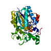
| Structure viewer | Molecule: MolmilJmol/JSmol |
|---|
- Downloads & links
-Download
| PDBx/mmCIF format | 5fml.cif.gz | 61 KB | Display | PDBx/mmCIF format |
|---|---|---|---|---|
| PDB format | pdb5fml.ent.gz | 44.6 KB | Display | PDB format |
| PDBx/mmJSON format | 5fml.json.gz | Tree view | PDBx/mmJSON format | |
| Others |  Other downloads Other downloads |
-Validation report
| Arichive directory | https://data.pdbj.org/pub/pdb/validation_reports/fm/5fmlftp://data.pdbj.org/pub/pdb/validation_reports/fm/5fml | HTTPS FTP |
|---|
-Related structure data
| Related structure data | 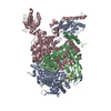 5epiC  5fmmC  5fmqC  5fmzC  4wrtS C: citing same article ( S: Starting model for refinement |
|---|---|
| Similar structure data |


-Links
 PDBj
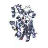
PDBj- Assembly
| Deposited unit | 
| ||||||||
|---|---|---|---|---|---|---|---|---|---|
| 1 |
| ||||||||
| Unit cell |
|
-Components
| #1: Protein/peptide | Mass: 3318.807 Da / Num. of mol.: 1 / Fragment: NLS PEPTIDE RESIDUES 742-770 / Source method: obtained synthetically / Source: (synth.) INFLUENZA B VIRUS / References: UniProt: Q5V8X3 | ||||||||
|---|---|---|---|---|---|---|---|---|---|
| #2: Protein | Mass: 23333.469 Da / Num. of mol.: 1 / Fragment: ENDONUCLEASE DOMAIN RESIDUES 1-197 Source method: isolated from a genetically manipulated source Source: (gene. exp.) INFLUENZA B VIRUS / Strain: B/MEMPHIS/13/2003/ / Plasmid: PETM11 / Production host:  | ||||||||
| #3: Chemical |   Mass: 24.305 Da / Num. of mol.: 2 / Source method: obtained synthetically / Formula: Mg Mass: 24.305 Da / Num. of mol.: 2 / Source method: obtained synthetically / Formula: Mg#4: Chemical | ChemComp-GOL / |   Mass: 92.094 Da / Num. of mol.: 1 / Source method: obtained synthetically / Formula: C3H8O3 Mass: 92.094 Da / Num. of mol.: 1 / Source method: obtained synthetically / Formula: C3H8O3#5: Water | ChemComp-HOH / |  Mass: 18.015 Da / Num. of mol.: 101 / Source method: isolated from a natural source / Formula: H2O Mass: 18.015 Da / Num. of mol.: 101 / Source method: isolated from a natural source / Formula: H2ONonpolymer details | GLYCEROL (GOL): CRYOPROTEC | Sequence details | ADDITIONAL | |
-Experimental details
-Experiment
| Experiment | Method: X-RAY DIFFRACTION / Number of used crystals: 1 |
|---|
- Sample preparation
| Crystal | Density Matthews: 2.7 Å3/Da / Density % sol: 54.3 % / Description: NONE |
|---|---|
| Crystal grow | pH: 6.5 / Details: 0.1 M MES PH 6.5, 25% PEG6000 |
-Data collection
| Diffraction | Mean temperature: 100 K |
|---|---|
| Diffraction source | Source: SYNCHROTRON / Site: ESRF  / Beamline: MASSIF-1 / Wavelength: 0.965 / Beamline: MASSIF-1 / Wavelength: 0.965 |
| Detector | Type: DECTRIS PILATUS 2M / Detector: PIXEL / Date: May 4, 2015 |
| Radiation | Protocol: SINGLE WAVELENGTH / Monochromatic (M) / Laue (L): M / Scattering type: x-ray |
| Radiation wavelength | Wavelength: 0.965 Å / Relative weight: 1 |
| Reflection | Resolution: 1.7→50 Å / Num. obs: 22279 / % possible obs: 98.2 % / Observed criterion σ(I): 0 / Redundancy: 2.72 % / Rmerge(I) obs: 0.03 / Net I/σ(I): 12.68 |
| Reflection shell | Resolution: 1.7→1.76 Å / Redundancy: 2.71 % / Rmerge(I) obs: 0.56 / Mean I/σ(I) obs: 1.84 / % possible all: 93 |
- Processing
| Software |
| ||||||||||||||||||||||||||||||||||||||||||||||||||||||||||||||||||||||||||||||||||||||||||||||||||||||||||||||||||||||||||||||||||||||||||||||||||||||||||||||||||||||||||||||||||||||
|---|---|---|---|---|---|---|---|---|---|---|---|---|---|---|---|---|---|---|---|---|---|---|---|---|---|---|---|---|---|---|---|---|---|---|---|---|---|---|---|---|---|---|---|---|---|---|---|---|---|---|---|---|---|---|---|---|---|---|---|---|---|---|---|---|---|---|---|---|---|---|---|---|---|---|---|---|---|---|---|---|---|---|---|---|---|---|---|---|---|---|---|---|---|---|---|---|---|---|---|---|---|---|---|---|---|---|---|---|---|---|---|---|---|---|---|---|---|---|---|---|---|---|---|---|---|---|---|---|---|---|---|---|---|---|---|---|---|---|---|---|---|---|---|---|---|---|---|---|---|---|---|---|---|---|---|---|---|---|---|---|---|---|---|---|---|---|---|---|---|---|---|---|---|---|---|---|---|---|---|---|---|---|---|
| Refinement | Method to determine structure: MOLECULAR REPLACEMENT Starting model: PDB ENTRY 4WRT Resolution: 1.7→40.42 Å / Cor.coef. Fo:Fc: 0.952 / Cor.coef. Fo:Fc free: 0.929 / SU B: 3.671 / SU ML: 0.119 / Cross valid method: THROUGHOUT / ESU R: 0.148 / ESU R Free: 0.146 / Stereochemistry target values: MAXIMUM LIKELIHOOD Details: HYDROGENS HAVE BEEN ADDED IN THE RIDING POSITIONS. U VALUES REFINED INDIVIDUALLY
| ||||||||||||||||||||||||||||||||||||||||||||||||||||||||||||||||||||||||||||||||||||||||||||||||||||||||||||||||||||||||||||||||||||||||||||||||||||||||||||||||||||||||||||||||||||||
| Solvent computation | Ion probe radii: 0.8 Å / Shrinkage radii: 0.8 Å / VDW probe radii: 1.2 Å / Solvent model: MASK | ||||||||||||||||||||||||||||||||||||||||||||||||||||||||||||||||||||||||||||||||||||||||||||||||||||||||||||||||||||||||||||||||||||||||||||||||||||||||||||||||||||||||||||||||||||||
| Displacement parameters | Biso mean: 32.895 Å2
| ||||||||||||||||||||||||||||||||||||||||||||||||||||||||||||||||||||||||||||||||||||||||||||||||||||||||||||||||||||||||||||||||||||||||||||||||||||||||||||||||||||||||||||||||||||||
| Refinement step | Cycle: LAST / Resolution: 1.7→40.42 Å
| ||||||||||||||||||||||||||||||||||||||||||||||||||||||||||||||||||||||||||||||||||||||||||||||||||||||||||||||||||||||||||||||||||||||||||||||||||||||||||||||||||||||||||||||||||||||
| Refine LS restraints |
|