 Movie
Movie Controller

[English] 日本語
 Yorodumi
- PDB-1z0k: Structure of GTP-Bound Rab4Q67L GTPase in complex with the centra... -
+ Open data

- Basic information
| Entry | Database: PDB / ID: 1z0k | ||||||
|---|---|---|---|---|---|---|---|
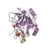
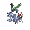
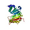
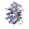
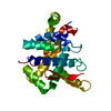
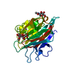
| Title | Structure of GTP-Bound Rab4Q67L GTPase in complex with the central Rab binding domain of Rabenosyn-5 | ||||||
 Components Components |
| ||||||
 Keywords Keywords | PROTEIN TRANSPORT / Rab GTPases / Rab4 / Rabenosyn / effector complex / vesicular trafficking | ||||||
| Function / homology |  Function and homology information Function and homology informationneurotransmitter receptor transport postsynaptic membrane to endosome / regulation of Golgi organization / Rab protein signal transduction / early endosome to Golgi transport / vesicle-mediated transport in synapse / Toll Like Receptor 9 (TLR9) Cascade / insulin-responsive compartment / Golgi to lysosome transport / RAB geranylgeranylation / phosphatidylinositol-3-phosphate binding ...neurotransmitter receptor transport postsynaptic membrane to endosome / regulation of Golgi organization / Rab protein signal transduction / early endosome to Golgi transport / vesicle-mediated transport in synapse / Toll Like Receptor 9 (TLR9) Cascade / insulin-responsive compartment / Golgi to lysosome transport / RAB geranylgeranylation / phosphatidylinositol-3-phosphate binding / MET receptor recycling / TBC/RABGAPs / antigen processing and presentation / endosomal transport / Synthesis of PIPs at the plasma membrane / regulation of endocytosis / vesicle-mediated transport / cytoplasmic vesicle membrane / small monomeric GTPase / Translocation of SLC2A4 (GLUT4) to the plasma membrane / recycling endosome / small GTPase binding / recycling endosome membrane / GDP binding / protein transport / Factors involved in megakaryocyte development and platelet production / G protein activity / early endosome membrane / vesicle / early endosome / endosome / endosome membrane / GTPase activity / GTP binding / perinuclear region of cytoplasm / glutamatergic synapse / extracellular exosome / zinc ion binding / metal ion binding / plasma membrane / cytosol Similarity search - Function | ||||||
| Biological species |  Homo sapiens (human) Homo sapiens (human) | ||||||
| Method |  X-RAY DIFFRACTION / MOLECULAR REPLACEMENT / Resolution: 1.92 Å X-RAY DIFFRACTION / MOLECULAR REPLACEMENT / Resolution: 1.92 Å | ||||||
 Authors Authors | Eathiraj, S. / Pan, X. / Ritacco, C. / Lambright, D.G. | ||||||
 Citation Citation | Journal: Nature / Year: 2005 Title: Structural basis of family-wide Rab GTPase recognition by rabenosyn-5. Authors: Eathiraj, S. / Pan, X. / Ritacco, C. / Lambright, D.G. | ||||||
| History |
|
- Structure visualization
| Structure viewer | Molecule: MolmilJmol/JSmol |
|---|
- Downloads & links
-Download
| PDBx/mmCIF format | 1z0k.cif.gz | 116.8 KB | Display | PDBx/mmCIF format |
|---|---|---|---|---|
| PDB format | pdb1z0k.ent.gz | 88.3 KB | Display | PDB format |
| PDBx/mmJSON format | 1z0k.json.gz | Tree view | PDBx/mmJSON format | |
| Others |  Other downloads Other downloads |
-Validation report
| Arichive directory | https://data.pdbj.org/pub/pdb/validation_reports/z0/1z0kftp://data.pdbj.org/pub/pdb/validation_reports/z0/1z0k | HTTPS FTP |
|---|
-Related structure data
| Related structure data | 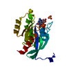 1yu9C 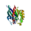 1yvdC 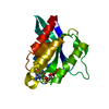 1yzkC 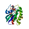 1yzlC  1yzmC  1yznC  1yzqC  1yztC 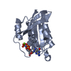 1yzuC 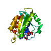 1z06C 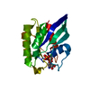 1z07C 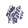 1z08C  1z0aC  1z0dC  1z0fC  1z0iC 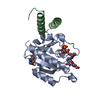 1z0jC 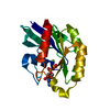 1z22C 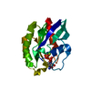 1z2aC C: citing same article ( |
|---|---|
| Similar structure data |


-Links
 PDBj
PDBj


- Assembly
| Deposited unit | 
| ||||||||
|---|---|---|---|---|---|---|---|---|---|
| 1 | 
| ||||||||
| 2 | 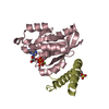
| ||||||||
| Unit cell |
|
-Components
-Protein , 2 types, 4 molecules ACBD
| #1: Protein | Mass: 19392.014 Da / Num. of mol.: 2 Source method: isolated from a genetically manipulated source Source: (gene. exp.) Homo sapiens (human) / Gene: RAB4 / Plasmid: modified pET28a / Production host:  #2: Protein | Mass: 7716.377 Da / Num. of mol.: 2 Source method: isolated from a genetically manipulated source Source: (gene. exp.) Homo sapiens (human) / Plasmid: pGEX6P1 / Production host: |
|---|
-Non-polymers , 4 types, 462 molecules 


| #3: Chemical |  Mass: 24.305 Da / Num. of mol.: 2 / Source method: obtained synthetically / Formula: Mg Mass: 24.305 Da / Num. of mol.: 2 / Source method: obtained synthetically / Formula: Mg#4: Chemical |  Mass: 523.180 Da / Num. of mol.: 2 / Source method: obtained synthetically / Formula: C10H16N5O14P3 / Comment: GTP, energy-carrying molecule*YM Mass: 523.180 Da / Num. of mol.: 2 / Source method: obtained synthetically / Formula: C10H16N5O14P3 / Comment: GTP, energy-carrying molecule*YM#5: Chemical | ChemComp-MES / |  Mass: 195.237 Da / Num. of mol.: 1 / Source method: obtained synthetically / Formula: C6H13NO4S / Comment: pH buffer*YM Mass: 195.237 Da / Num. of mol.: 1 / Source method: obtained synthetically / Formula: C6H13NO4S / Comment: pH buffer*YM#6: Water | ChemComp-HOH / | Mass: 18.015 Da / Num. of mol.: 457 / Source method: isolated from a natural source / Formula: H2O |
|---|
-Experimental details
-Experiment
| Experiment | Method: X-RAY DIFFRACTION / Number of used crystals: 1 |
|---|
- Sample preparation
| Crystal | Density Matthews: 2.58 Å3/Da / Density % sol: 51.91 % |
|---|---|
| Crystal grow | Temperature: 291 K / Method: vapor diffusion, hanging drop / pH: 6 Details: 20% PEG 4000, 200mM ammonium fluoride, 50mM MES, pH 6.0, VAPOR DIFFUSION, HANGING DROP, temperature 291K |
-Data collection
| Diffraction | Mean temperature: 100 K |
|---|---|
| Diffraction source | Source: ROTATING ANODE / Type: RIGAKU RUH3R / Wavelength: 1.5418 |
| Detector | Type: MAR scanner 300 mm plate / Detector: IMAGE PLATE / Date: May 11, 2004 / Details: osmic mirror |
| Radiation | Protocol: SINGLE WAVELENGTH / Monochromatic (M) / Laue (L): M / Scattering type: x-ray |
| Radiation wavelength | Wavelength: 1.5418 Å / Relative weight: 1 |
| Reflection | Resolution: 1.9→50 Å / Num. obs: 42193 / % possible obs: 99.6 % / Observed criterion σ(F): 0 / Observed criterion σ(I): 0 / Redundancy: 5 % / Biso Wilson estimate: 41.7 Å2 / Rsym value: 0.054 / Net I/σ(I): 27.29 |
| Reflection shell | Resolution: 1.9→1.96 Å / Redundancy: 5 % / Mean I/σ(I) obs: 4 / Num. unique all: 3439 / Rsym value: 0.379 / % possible all: 98.9 |
- Processing
| Software |
| ||||||||||||||||||||||||||||||||||||||||||||||||||||||||||||||||||||||||||||||||||||||||||||||||||||||||||||||||||||||||||||||||||||||||||||||||||||||||||||||||||||||||||
|---|---|---|---|---|---|---|---|---|---|---|---|---|---|---|---|---|---|---|---|---|---|---|---|---|---|---|---|---|---|---|---|---|---|---|---|---|---|---|---|---|---|---|---|---|---|---|---|---|---|---|---|---|---|---|---|---|---|---|---|---|---|---|---|---|---|---|---|---|---|---|---|---|---|---|---|---|---|---|---|---|---|---|---|---|---|---|---|---|---|---|---|---|---|---|---|---|---|---|---|---|---|---|---|---|---|---|---|---|---|---|---|---|---|---|---|---|---|---|---|---|---|---|---|---|---|---|---|---|---|---|---|---|---|---|---|---|---|---|---|---|---|---|---|---|---|---|---|---|---|---|---|---|---|---|---|---|---|---|---|---|---|---|---|---|---|---|---|---|---|---|---|
| Refinement | Method to determine structure: MOLECULAR REPLACEMENT Starting model: Rab4 GTPase Resolution: 1.92→15 Å / Cor.coef. Fo:Fc: 0.949 / Cor.coef. Fo:Fc free: 0.934 / SU B: 4.5 / SU ML: 0.13 / Cross valid method: THROUGHOUT / σ(F): 0 / σ(I): 0 / ESU R: 0.188 / ESU R Free: 0.165 / Stereochemistry target values: MAXIMUM LIKELIHOOD / Details: HYDROGENS HAVE BEEN ADDED IN THE RIDING POSITIONS
| ||||||||||||||||||||||||||||||||||||||||||||||||||||||||||||||||||||||||||||||||||||||||||||||||||||||||||||||||||||||||||||||||||||||||||||||||||||||||||||||||||||||||||
| Solvent computation | Ion probe radii: 0.8 Å / Shrinkage radii: 0.8 Å / VDW probe radii: 1.2 Å / Solvent model: MASK | ||||||||||||||||||||||||||||||||||||||||||||||||||||||||||||||||||||||||||||||||||||||||||||||||||||||||||||||||||||||||||||||||||||||||||||||||||||||||||||||||||||||||||
| Displacement parameters | Biso mean: 33.045 Å2
| ||||||||||||||||||||||||||||||||||||||||||||||||||||||||||||||||||||||||||||||||||||||||||||||||||||||||||||||||||||||||||||||||||||||||||||||||||||||||||||||||||||||||||
| Refinement step | Cycle: LAST / Resolution: 1.92→15 Å
| ||||||||||||||||||||||||||||||||||||||||||||||||||||||||||||||||||||||||||||||||||||||||||||||||||||||||||||||||||||||||||||||||||||||||||||||||||||||||||||||||||||||||||
| Refine LS restraints |
| ||||||||||||||||||||||||||||||||||||||||||||||||||||||||||||||||||||||||||||||||||||||||||||||||||||||||||||||||||||||||||||||||||||||||||||||||||||||||||||||||||||||||||
| LS refinement shell | Resolution: 1.92→1.969 Å / Total num. of bins used: 20
|
