 Movie
Movie Controller

[English] 日本語
 Yorodumi
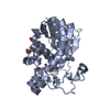
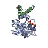
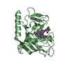
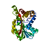
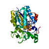
- PDB-2hls: The crystal structure of a protein disulfide oxidoreductase from ... -
+ Open data

- Basic information
| Entry | Database: PDB / ID: 2hls | ||||||
|---|---|---|---|---|---|---|---|
| Title | The crystal structure of a protein disulfide oxidoreductase from Aeropyrum pernix k1 | ||||||
 Components Components | protein disulfide oxidoreductase | ||||||
 Keywords Keywords | OXIDOREDUCTASE / protein disulfide oxidoreductase / thioredoxin fold | ||||||
| Function / homology | Glutaredoxin-like, bacteria/archaea / Thioredoxin domain / Thioredoxin-like fold / Glutaredoxin / Glutaredoxin / Thioredoxin-like superfamily / 3-Layer(aba) Sandwich / Alpha Beta / Protein-disulfide oxidoreductase Function and homology information Function and homology information | ||||||
| Biological species |   Aeropyrum pernix (archaea) Aeropyrum pernix (archaea) | ||||||
| Method |  X-RAY DIFFRACTION / SYNCHROTRON / MOLECULAR REPLACEMENT / Resolution: 1.93 Å X-RAY DIFFRACTION / SYNCHROTRON / MOLECULAR REPLACEMENT / Resolution: 1.93 Å | ||||||
 Authors Authors | D'Ambrosio, K. / De Simone, G. | ||||||
 Citation Citation | Journal: J.Mol.Biol. / Year: 2006 Title: A Novel Member of the Protein Disulfide Oxidoreductase Family from Aeropyrum pernix K1: Structure, Function and Electrostatics. Authors: D'Ambrosio, K. / Pedone, E. / Langella, E. / De Simone, G. / Rossi, M. / Pedone, C. / Bartolucci, S. | ||||||
| History |
|
- Structure visualization
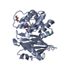
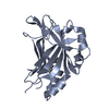
| Structure viewer | Molecule: MolmilJmol/JSmol |
|---|
- Downloads & links
-Download
| PDBx/mmCIF format | 2hls.cif.gz | 112 KB | Display | PDBx/mmCIF format |
|---|---|---|---|---|
| PDB format | pdb2hls.ent.gz | 86.3 KB | Display | PDB format |
| PDBx/mmJSON format | 2hls.json.gz | Tree view | PDBx/mmJSON format | |
| Others |  Other downloads Other downloads |
-Validation report
| Arichive directory | https://data.pdbj.org/pub/pdb/validation_reports/hl/2hlsftp://data.pdbj.org/pub/pdb/validation_reports/hl/2hls | HTTPS FTP |
|---|
-Related structure data
| Related structure data |  2aytS S: Starting model for refinement |
|---|---|
| Similar structure data |

-Links
 PDBj
PDBj- Assembly
| Deposited unit | 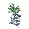
| ||||||||
|---|---|---|---|---|---|---|---|---|---|
| 1 | 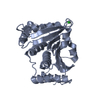
| ||||||||
| 2 | 
| ||||||||
| 3 | 
| ||||||||
| Unit cell |
| ||||||||
| Details | The biological assembly is a monomer |
-Components
| #1: Protein | Mass: 27364.143 Da / Num. of mol.: 2 Source method: isolated from a genetically manipulated source Source: (gene. exp.) Aeropyrum pernix (archaea) / Strain: K1 / Plasmid: pTrc99A / Production host:  #2: Chemical |   Mass: 35.453 Da / Num. of mol.: 2 / Source method: obtained synthetically / Formula: Cl Mass: 35.453 Da / Num. of mol.: 2 / Source method: obtained synthetically / Formula: Cl#3: Water | ChemComp-HOH / |  Mass: 18.015 Da / Num. of mol.: 424 / Source method: isolated from a natural source / Formula: H2O Mass: 18.015 Da / Num. of mol.: 424 / Source method: isolated from a natural source / Formula: H2OHas protein modification | Y | |
|---|
-Experimental details
-Experiment
| Experiment | Method: X-RAY DIFFRACTION / Number of used crystals: 1 |
|---|
- Sample preparation
| Crystal | Density Matthews: 2.71 Å3/Da / Density % sol: 54.54 % |
|---|---|
| Crystal grow | Temperature: 298 K / Method: vapor diffusion, hanging drop / pH: 8 Details: 2M ammonium sulfate, 2% PEG 400, 0.1M HEPES, pH 8, VAPOR DIFFUSION, HANGING DROP, temperature 298K |
-Data collection
| Diffraction | Mean temperature: 100 K |
|---|---|
| Diffraction source | Source: SYNCHROTRON / Site: ELETTRA  / Beamline: 5.2R / Wavelength: 1.2 Å / Beamline: 5.2R / Wavelength: 1.2 Å |
| Detector | Type: MAR CCD 165 mm / Detector: CCD / Date: Dec 14, 2004 |
| Radiation | Protocol: SINGLE WAVELENGTH / Monochromatic (M) / Laue (L): M / Scattering type: x-ray |
| Radiation wavelength | Wavelength: 1.2 Å / Relative weight: 1 |
| Reflection | Resolution: 1.93→20 Å / Num. obs: 44495 / % possible obs: 98.1 % / Rsym value: 0.034 / Net I/σ(I): 36.9 |
| Reflection shell | Resolution: 1.93→2 Å / Mean I/σ(I) obs: 2.9 / Rsym value: 0.297 / % possible all: 87.5 |
- Processing
| Software |
| ||||||||||||||||||||
|---|---|---|---|---|---|---|---|---|---|---|---|---|---|---|---|---|---|---|---|---|---|
| Refinement | Method to determine structure: MOLECULAR REPLACEMENT Starting model: pdb entry 2AYT Resolution: 1.93→20 Å / σ(F): 0 / Stereochemistry target values: Engh & Huber
| ||||||||||||||||||||
| Refinement step | Cycle: LAST / Resolution: 1.93→20 Å
| ||||||||||||||||||||
| Refine LS restraints |
|
