 Movie
Movie Controller

[English] 日本語
 Yorodumi
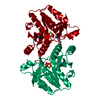
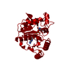
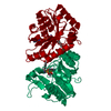
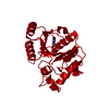
- PDB-1byi: STRUCTURE OF APO-DETHIOBIOTIN SYNTHASE AT 0.97 ANGSTROMS RESOLUTION -
+ Open data

- Basic information
| Entry | Database: PDB / ID: 1byi | ||||||
|---|---|---|---|---|---|---|---|
| Title | STRUCTURE OF APO-DETHIOBIOTIN SYNTHASE AT 0.97 ANGSTROMS RESOLUTION | ||||||
 Components Components | DETHIOBIOTIN SYNTHASE | ||||||
 Keywords Keywords | LIGASE / BIOTIN SYNTHESIS / CYCLO-LIGASE | ||||||
| Function / homology |  Function and homology information Function and homology informationdethiobiotin synthase / dethiobiotin synthase activity / biotin biosynthetic process / magnesium ion binding / protein homodimerization activity / ATP binding / cytosol Similarity search - Function | ||||||
| Biological species |  | ||||||
| Method |  X-RAY DIFFRACTION / SYNCHROTRON / MOLECULAR REPLACEMENT / Resolution: 0.97 Å X-RAY DIFFRACTION / SYNCHROTRON / MOLECULAR REPLACEMENT / Resolution: 0.97 Å | ||||||
 Authors Authors | Sandalova, T. / Schneider, G. / Kaeck, H. / Lindqvist, Y. | ||||||
 Citation Citation | Journal: Acta Crystallogr.,Sect.D / Year: 1999 Title: Structure of dethiobiotin synthetase at 0.97 A resolution. Authors: Sandalova, T. / Schneider, G. / Kack, H. / Lindqvist, Y. #1: Journal: Structure / Year: 1994Title: Crystal Structure of an ATP-Dependent Carboxylase, Dethiobiotin Synthetase, at 1.65 A Resolution Authors: Huang, W. / Lindqvist, Y. / Schneider, G. / Gibson, K.J. / Flint, D. / Lorimer, G. | ||||||
| History |
|
- Structure visualization
| Structure viewer | Molecule: MolmilJmol/JSmol |
|---|
- Downloads & links
-Download
| PDBx/mmCIF format | 1byi.cif.gz | 120.4 KB | Display | PDBx/mmCIF format |
|---|---|---|---|---|
| PDB format | pdb1byi.ent.gz | 94.5 KB | Display | PDB format |
| PDBx/mmJSON format | 1byi.json.gz | Tree view | PDBx/mmJSON format | |
| Others |  Other downloads Other downloads |
-Validation report
| Arichive directory | https://data.pdbj.org/pub/pdb/validation_reports/by/1byiftp://data.pdbj.org/pub/pdb/validation_reports/by/1byi | HTTPS FTP |
|---|
-Related structure data
| Related structure data | 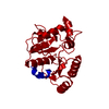 1dtsS S: Starting model for refinement |
|---|---|
| Similar structure data |


-Links
 PDBj
PDBj- Assembly
| Deposited unit | 
| ||||||||||||||||||
|---|---|---|---|---|---|---|---|---|---|---|---|---|---|---|---|---|---|---|---|
| 1 | 
| ||||||||||||||||||
| Unit cell |
| ||||||||||||||||||
| Components on special symmetry positions |
|
-Components
| #1: Protein | Mass: 24028.289 Da / Num. of mol.: 1 / Source method: isolated from a natural source / Source: (natural) |
|---|---|
| #2: Water | ChemComp-HOH /  Mass: 18.015 Da / Num. of mol.: 403 / Source method: isolated from a natural source / Formula: H2O Mass: 18.015 Da / Num. of mol.: 403 / Source method: isolated from a natural source / Formula: H2O |
-Experimental details
-Experiment
| Experiment | Method: X-RAY DIFFRACTION / Number of used crystals: 1 |
|---|
- Sample preparation
| Crystal | Density Matthews: 2 Å3/Da / Density % sol: 38 % | |||||||||||||||||||||||||
|---|---|---|---|---|---|---|---|---|---|---|---|---|---|---|---|---|---|---|---|---|---|---|---|---|---|---|
| Crystal grow | pH: 6.5 / Details: pH 6.5 | |||||||||||||||||||||||||
| Crystal grow | *PLUS pH: 6 / Method: microdialysis | |||||||||||||||||||||||||
| Components of the solutions | *PLUS
|
-Data collection
| Diffraction | Mean temperature: 110 K |
|---|---|
| Diffraction source | Source: SYNCHROTRON / Site: EMBL/DESY, HAMBURG  / Beamline: X11 / Wavelength: 0.9082 / Beamline: X11 / Wavelength: 0.9082 |
| Detector | Type: MAR scanner 300 mm plate / Detector: IMAGE PLATE / Date: Dec 1, 1996 |
| Radiation | Monochromatic (M) / Laue (L): M / Scattering type: x-ray |
| Radiation wavelength | Wavelength: 0.9082 Å / Relative weight: 1 |
| Reflection | Resolution: 0.97→15 Å / Num. obs: 116232 / % possible obs: 98.5 % / Observed criterion σ(I): 0 / Redundancy: 4.2 % / Rmerge(I) obs: 0.025 / Net I/σ(I): 54.4 |
| Reflection shell | Resolution: 0.97→0.98 Å / Redundancy: 2.1 % / Mean I/σ(I) obs: 4.2 / Rsym value: 0.245 / % possible all: 94.3 |
| Reflection | *PLUS Num. measured all: 494742 |
| Reflection shell | *PLUS % possible obs: 94.3 % / Rmerge(I) obs: 0.245 |
- Processing
| Software |
| |||||||||||||||||||||||||||||||||
|---|---|---|---|---|---|---|---|---|---|---|---|---|---|---|---|---|---|---|---|---|---|---|---|---|---|---|---|---|---|---|---|---|---|---|
| Refinement | Method to determine structure: MOLECULAR REPLACEMENT Starting model: PDB ENTRY 1DTS Resolution: 0.97→15 Å / Num. parameters: 20493 / Num. restraintsaints: 25635 / σ(F): 0 / Stereochemistry target values: ENGH AND HUBER
| |||||||||||||||||||||||||||||||||
| Solvent computation | Solvent model: MOEWS & KRETSINGER | |||||||||||||||||||||||||||||||||
| Refine analyze | Num. disordered residues: 79 / Occupancy sum hydrogen: 1684.4 / Occupancy sum non hydrogen: 2075 | |||||||||||||||||||||||||||||||||
| Refinement step | Cycle: LAST / Resolution: 0.97→15 Å
| |||||||||||||||||||||||||||||||||
| Refine LS restraints |
| |||||||||||||||||||||||||||||||||
| Software | *PLUS Name: SHELXL-97 / Classification: refinement | |||||||||||||||||||||||||||||||||
| Refine LS restraints | *PLUS Type: s_chiral_restr / Dev ideal: 0.078 |
