 ムービー
ムービー コントローラー


+ データを開く

- 基本情報
基本情報
| 登録情報 | データベース: PDB / ID: 5nna | ||||||
|---|---|---|---|---|---|---|---|
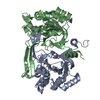
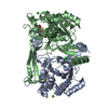
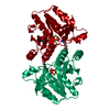
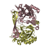
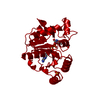
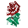
| タイトル | Isatin hydrolase A (IHA) from Labrenzia aggregata bound to benzyl benzoate | ||||||
 要素 要素 | isatin hydrolase A | ||||||
 キーワード キーワード | HYDROLASE / Isatin / Labrenzia aggregata / benzyl benzoate | ||||||
| 機能・相同性 |  機能・相同性情報 機能・相同性情報 | ||||||
| 生物種 |  Labrenzia aggregata (バクテリア) Labrenzia aggregata (バクテリア) | ||||||
| 手法 |  X線回折 / シンクロトロン / 分子置換 / 解像度: 1.5 Å X線回折 / シンクロトロン / 分子置換 / 解像度: 1.5 Å | ||||||
 データ登録者 データ登録者 | Sommer, T. / Bjerregaard-Andersen, K. / Morth, J.P. | ||||||
 引用 引用 | ジャーナル: Sci Rep / 年: 2018 タイトル: A fundamental catalytic difference between zinc and manganese dependent enzymes revealed in a bacterial isatin hydrolase. 著者: Sommer, T. / Bjerregaard-Andersen, K. / Uribe, L. / Etzerodt, M. / Diezemann, G. / Gauss, J. / Cascella, M. / Morth, J.P. | ||||||
| 履歴 |
|
- 構造の表示
構造の表示
| 構造ビューア | 分子: MolmilJmol/JSmol |
|---|
- ダウンロードとリンク
-ダウンロード
| PDBx/mmCIF形式 | 5nna.cif.gz | 237.7 KB | 表示 | PDBx/mmCIF形式 |
|---|---|---|---|---|
| PDB形式 | pdb5nna.ent.gz | 189.2 KB | 表示 | PDB形式 |
| PDBx/mmJSON形式 | 5nna.json.gz | ツリー表示 | PDBx/mmJSON形式 | |
| その他 |  その他のダウンロード その他のダウンロード |
-検証レポート
| アーカイブディレクトリ | https://data.pdbj.org/pub/pdb/validation_reports/nn/5nnaftp://data.pdbj.org/pub/pdb/validation_reports/nn/5nna | HTTPS FTP |
|---|
-関連構造データ


-リンク
 PDBj
PDBj- 集合体
集合体
| 登録構造単位 | 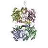
| ||||||||
|---|---|---|---|---|---|---|---|---|---|
| 1 | 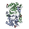
| ||||||||
| 2 | 
| ||||||||
| 単位格子 |
|
-要素
-タンパク質 , 1種, 4分子 ABCD
| #1: タンパク質 | 分子量: 28810.295 Da / 分子数: 4 / 由来タイプ: 組換発現 詳細: The His-tag was visible in the density in this molecule 由来: (組換発現) Labrenzia aggregata (バクテリア)遺伝子: SIAM614_30646 / 発現宿主: |
|---|
-非ポリマー , 6種, 1032分子 


| #2: 化合物 | ChemComp-BZM /  分子量: 212.244 Da / 分子数: 4 / 由来タイプ: 合成 / 式: C14H12O2 / コメント: 薬剤*YM 分子量: 212.244 Da / 分子数: 4 / 由来タイプ: 合成 / 式: C14H12O2 / コメント: 薬剤*YM#3: 化合物 | ChemComp-MN /  分子量: 54.938 Da / 分子数: 4 / 由来タイプ: 合成 / 式: Mn 分子量: 54.938 Da / 分子数: 4 / 由来タイプ: 合成 / 式: Mn#4: 化合物 | ChemComp-BTB / |  分子量: 209.240 Da / 分子数: 1 / 由来タイプ: 合成 / 式: C8H19NO5 / コメント: pH緩衝剤*YM 分子量: 209.240 Da / 分子数: 1 / 由来タイプ: 合成 / 式: C8H19NO5 / コメント: pH緩衝剤*YM#5: 化合物 | ChemComp-1PE / |  分子量: 238.278 Da / 分子数: 1 / 由来タイプ: 合成 / 式: C10H22O6 / コメント: 沈殿剤*YM 分子量: 238.278 Da / 分子数: 1 / 由来タイプ: 合成 / 式: C10H22O6 / コメント: 沈殿剤*YM#6: 化合物 | ChemComp-PEG / |  分子量: 106.120 Da / 分子数: 1 / 由来タイプ: 天然 / 式: C4H10O3 分子量: 106.120 Da / 分子数: 1 / 由来タイプ: 天然 / 式: C4H10O3#7: 水 | ChemComp-HOH / | 分子量: 18.015 Da / 分子数: 1021 / 由来タイプ: 天然 / 式: H2O |
|---|
-実験情報
-実験
| 実験 | 手法: X線回折 / 使用した結晶の数: 1 |
|---|
- 試料調製
試料調製
| 結晶 | マシュー密度: 2.02 Å3/Da / 溶媒含有率: 39.12 % |
|---|---|
| 結晶化 | 温度: 295 K / 手法: 蒸気拡散法, ハンギングドロップ法 / pH: 7.5 / 詳細: 28 % PEG 1500 and 1 mM MnCl2 |
-データ収集
| 回折 | 平均測定温度: 100 K |
|---|---|
| 放射光源 | 由来: シンクロトロン / サイト: Diamond  / ビームライン: I02 / 波長: 1.0004 Å / ビームライン: I02 / 波長: 1.0004 Å |
| 検出器 | タイプ: DECTRIS PILATUS3 S 6M / 検出器: PIXEL / 日付: 2015年4月1日 |
| 放射 | プロトコル: SINGLE WAVELENGTH / 単色(M)・ラウエ(L): M / 散乱光タイプ: x-ray |
| 放射波長 | 波長: 1.0004 Å / 相対比: 1 |
| 反射 | 解像度: 1.5→63.544 Å / Num. obs: 135214 / % possible obs: 95.7 % / 冗長度: 3 % / Net I/σ(I): 10 |
| 反射 シェル | 解像度: 1.5→1.54 Å |
- 解析
解析
| ソフトウェア |
| |||||||||||||||||||||||||||||||||||||||||||||||||||||||||||||||||||||||||||||||||||||||||||||||||||||||||||||||||||||||||||||||||||||||||||||||||||||||||||||||||||||||||||||||||||||||||||||||||||||||||||||||||||||||||
|---|---|---|---|---|---|---|---|---|---|---|---|---|---|---|---|---|---|---|---|---|---|---|---|---|---|---|---|---|---|---|---|---|---|---|---|---|---|---|---|---|---|---|---|---|---|---|---|---|---|---|---|---|---|---|---|---|---|---|---|---|---|---|---|---|---|---|---|---|---|---|---|---|---|---|---|---|---|---|---|---|---|---|---|---|---|---|---|---|---|---|---|---|---|---|---|---|---|---|---|---|---|---|---|---|---|---|---|---|---|---|---|---|---|---|---|---|---|---|---|---|---|---|---|---|---|---|---|---|---|---|---|---|---|---|---|---|---|---|---|---|---|---|---|---|---|---|---|---|---|---|---|---|---|---|---|---|---|---|---|---|---|---|---|---|---|---|---|---|---|---|---|---|---|---|---|---|---|---|---|---|---|---|---|---|---|---|---|---|---|---|---|---|---|---|---|---|---|---|---|---|---|---|---|---|---|---|---|---|---|---|---|---|---|---|---|---|---|---|
| 精密化 | 構造決定の手法: 分子置換 開始モデル: 4M8D 解像度: 1.5→63.544 Å / SU ML: 0.14 / 交差検証法: FREE R-VALUE / σ(F): 1.98 / 位相誤差: 16.66
| |||||||||||||||||||||||||||||||||||||||||||||||||||||||||||||||||||||||||||||||||||||||||||||||||||||||||||||||||||||||||||||||||||||||||||||||||||||||||||||||||||||||||||||||||||||||||||||||||||||||||||||||||||||||||
| 溶媒の処理 | 減衰半径: 0.9 Å / VDWプローブ半径: 1.11 Å | |||||||||||||||||||||||||||||||||||||||||||||||||||||||||||||||||||||||||||||||||||||||||||||||||||||||||||||||||||||||||||||||||||||||||||||||||||||||||||||||||||||||||||||||||||||||||||||||||||||||||||||||||||||||||
| 精密化ステップ | サイクル: LAST / 解像度: 1.5→63.544 Å
| |||||||||||||||||||||||||||||||||||||||||||||||||||||||||||||||||||||||||||||||||||||||||||||||||||||||||||||||||||||||||||||||||||||||||||||||||||||||||||||||||||||||||||||||||||||||||||||||||||||||||||||||||||||||||
| 拘束条件 |
| |||||||||||||||||||||||||||||||||||||||||||||||||||||||||||||||||||||||||||||||||||||||||||||||||||||||||||||||||||||||||||||||||||||||||||||||||||||||||||||||||||||||||||||||||||||||||||||||||||||||||||||||||||||||||
| LS精密化 シェル |
|
