 Movie
Movie Controller

[English] 日本語
 Yorodumi
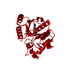
- PDB-1dak: DETHIOBIOTIN SYNTHETASE FROM ESCHERICHIA COLI, COMPLEX REACTION I... -
+ Open data

- Basic information
| Entry | Database: PDB / ID: 1dak | ||||||
|---|---|---|---|---|---|---|---|
| Title | DETHIOBIOTIN SYNTHETASE FROM ESCHERICHIA COLI, COMPLEX REACTION INTERMEDIATE ADP AND MIXED ANHYDRIDE | ||||||
 Components Components | DETHIOBIOTIN SYNTHETASE | ||||||
 Keywords Keywords | LIGASE / PHOSHPORYL TRANSFER / BIOTIN BIOSYNTHESIS / KINETIC CRYSTALLOGRAPHY | ||||||
| Function / homology |  Function and homology information Function and homology informationdethiobiotin synthase / dethiobiotin synthase activity / biotin biosynthetic process / magnesium ion binding / protein homodimerization activity / ATP binding / cytosol Similarity search - Function | ||||||
| Biological species |  | ||||||
| Method |  X-RAY DIFFRACTION / SYNCHROTRON / DIFFERENCE FOURIER / Resolution: 1.6 Å X-RAY DIFFRACTION / SYNCHROTRON / DIFFERENCE FOURIER / Resolution: 1.6 Å | ||||||
 Authors Authors | Kaeck, H. / Gibson, K.J. / Lindqvist, Y. / Schneider, G. | ||||||
 Citation Citation | Journal: Proc.Natl.Acad.Sci.USA / Year: 1998 Title: Snapshot of a phosphorylated substrate intermediate by kinetic crystallography. Authors: Kack, H. / Gibson, K.J. / Lindqvist, Y. / Schneider, G. | ||||||
| History |
|
- Structure visualization
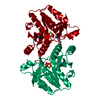
| Structure viewer | Molecule: MolmilJmol/JSmol |
|---|
- Downloads & links
-Download
| PDBx/mmCIF format | 1dak.cif.gz | 62.7 KB | Display | PDBx/mmCIF format |
|---|---|---|---|---|
| PDB format | pdb1dak.ent.gz | 44 KB | Display | PDB format |
| PDBx/mmJSON format | 1dak.json.gz | Tree view | PDBx/mmJSON format | |
| Others |  Other downloads Other downloads |
-Validation report
| Arichive directory | https://data.pdbj.org/pub/pdb/validation_reports/da/1dakftp://data.pdbj.org/pub/pdb/validation_reports/da/1dak | HTTPS FTP |
|---|
-Related structure data
| Related structure data | 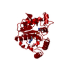 1a82C 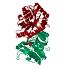 1dahS S: Starting model for refinement C: citing same article ( |
|---|---|
| Similar structure data |


-Links
 PDBj
PDBj- Assembly
| Deposited unit | 
| ||||||||
|---|---|---|---|---|---|---|---|---|---|
| 1 | 
| ||||||||
| Unit cell |
|
-Components
-Protein , 1 types, 1 molecules A
| #1: Protein | Mass: 24028.289 Da / Num. of mol.: 1 Source method: isolated from a genetically manipulated source Source: (gene. exp.) |
|---|
-Non-polymers , 5 types, 221 molecules 


| #2: Chemical | ChemComp-PO4 /  Mass: 94.971 Da / Num. of mol.: 1 / Source method: obtained synthetically / Formula: PO4 Mass: 94.971 Da / Num. of mol.: 1 / Source method: obtained synthetically / Formula: PO4 | ||||||
|---|---|---|---|---|---|---|---|
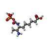
| #3: Chemical |  Mass: 24.305 Da / Num. of mol.: 2 / Source method: obtained synthetically / Formula: Mg Mass: 24.305 Da / Num. of mol.: 2 / Source method: obtained synthetically / Formula: Mg#4: Chemical | ChemComp-DPU / |  Mass: 312.257 Da / Num. of mol.: 1 / Source method: obtained synthetically / Formula: C10H21N2O7P Mass: 312.257 Da / Num. of mol.: 1 / Source method: obtained synthetically / Formula: C10H21N2O7P#5: Chemical | ChemComp-ADP / |  Mass: 427.201 Da / Num. of mol.: 1 / Source method: obtained synthetically / Formula: C10H15N5O10P2 / Comment: ADP, energy-carrying molecule*YM Mass: 427.201 Da / Num. of mol.: 1 / Source method: obtained synthetically / Formula: C10H15N5O10P2 / Comment: ADP, energy-carrying molecule*YM#6: Water | ChemComp-HOH / | Mass: 18.015 Da / Num. of mol.: 216 / Source method: isolated from a natural source / Formula: H2O |
-Experimental details
-Experiment
| Experiment | Method: X-RAY DIFFRACTION / Number of used crystals: 1 |
|---|
- Sample preparation
| Crystal | Density Matthews: 2.13 Å3/Da / Density % sol: 42.3 % | ||||||||||||||||||||||||||||||
|---|---|---|---|---|---|---|---|---|---|---|---|---|---|---|---|---|---|---|---|---|---|---|---|---|---|---|---|---|---|---|---|
| Crystal grow | pH: 8.5 / Details: pH 8.5 | ||||||||||||||||||||||||||||||
| Crystal grow | *PLUS pH: 6.5 / Method: vapor diffusionDetails: drop contained 0.003ml of protein solution mixed with 0.074ml of well solution. | ||||||||||||||||||||||||||||||
| Components of the solutions | *PLUS
|
-Data collection
| Diffraction | Mean temperature: 100 K |
|---|---|
| Diffraction source | Source: SYNCHROTRON / Site: ESRF  / Beamline: ID09 / Wavelength: 0.7606 / Beamline: ID09 / Wavelength: 0.7606 |
| Detector | Type: PRINCETON 2K / Detector: CCD / Date: Oct 1, 1997 |
| Radiation | Monochromatic (M) / Laue (L): M / Scattering type: x-ray |
| Radiation wavelength | Wavelength: 0.7606 Å / Relative weight: 1 |
| Reflection | Resolution: 1.6→25 Å / Num. obs: 86120 / % possible obs: 99 % / Redundancy: 3.2 % / Rsym value: 0.078 / Net I/σ(I): 16 |
| Reflection shell | Resolution: 1.6→1.66 Å / Redundancy: 2.6 % / Mean I/σ(I) obs: 2.8 / Rsym value: 0.43 / % possible all: 92.4 |
| Reflection | *PLUS Rmerge(I) obs: 0.078 |
| Reflection shell | *PLUS % possible obs: 92.4 % / Rmerge(I) obs: 0.43 |
- Processing
| Software |
| ||||||||||||||||||||||||||||||||||||||||||||||||||||||||||||||||||||||||||||||||||||
|---|---|---|---|---|---|---|---|---|---|---|---|---|---|---|---|---|---|---|---|---|---|---|---|---|---|---|---|---|---|---|---|---|---|---|---|---|---|---|---|---|---|---|---|---|---|---|---|---|---|---|---|---|---|---|---|---|---|---|---|---|---|---|---|---|---|---|---|---|---|---|---|---|---|---|---|---|---|---|---|---|---|---|---|---|---|
| Refinement | Method to determine structure: DIFFERENCE FOURIER Starting model: PDB ENTRY 1DAH, WITH NON-PROTEIN ATOMS EXCLUDED Resolution: 1.6→20 Å / Cross valid method: THROUGHOUT / σ(F): 0
| ||||||||||||||||||||||||||||||||||||||||||||||||||||||||||||||||||||||||||||||||||||
| Displacement parameters | Biso mean: 13.6 Å2 | ||||||||||||||||||||||||||||||||||||||||||||||||||||||||||||||||||||||||||||||||||||
| Refinement step | Cycle: LAST / Resolution: 1.6→20 Å
| ||||||||||||||||||||||||||||||||||||||||||||||||||||||||||||||||||||||||||||||||||||
| Refine LS restraints |
| ||||||||||||||||||||||||||||||||||||||||||||||||||||||||||||||||||||||||||||||||||||
| Software | *PLUS Name: REFMAC / Classification: refinement | ||||||||||||||||||||||||||||||||||||||||||||||||||||||||||||||||||||||||||||||||||||
| Refinement | *PLUS Rfactor obs: 0.184 | ||||||||||||||||||||||||||||||||||||||||||||||||||||||||||||||||||||||||||||||||||||
| Solvent computation | *PLUS | ||||||||||||||||||||||||||||||||||||||||||||||||||||||||||||||||||||||||||||||||||||
| Displacement parameters | *PLUS |
