 Movie
Movie Controller

[English] 日本語
 Yorodumi
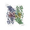
- PDB-5epi: CRYSTAL STRUCTURE OF INFLUENZA B POLYMERASE WITH BOUND 5' CRNA EX... -
+ Open data

- Basic information
| Entry | Database: PDB / ID: 5epi | ||||||
|---|---|---|---|---|---|---|---|
| Title | CRYSTAL STRUCTURE OF INFLUENZA B POLYMERASE WITH BOUND 5' CRNA EXHIBITS A NOVEL DOMAIN ARRANGEMENT | ||||||
 Components Components |
| ||||||
 Keywords Keywords | TRANSFERASE / INFLUENZA B VIRUS / RNA-DEPENDENT RNA POLYMERASE / HETEROTRIMER / SUBUNITS PA / PB1 / PB2 / VIRAL RNA / CRNA 5' END / INFLUENZA | ||||||
| Function / homology |  Function and homology information Function and homology informationcap snatching / viral transcription / symbiont-mediated suppression of host mRNA transcription via inhibition of RNA polymerase II activity / 7-methylguanosine mRNA capping / host cell mitochondrion / virion component / endonuclease activity / Hydrolases; Acting on ester bonds / host cell cytoplasm / symbiont-mediated suppression of host gene expression ...cap snatching / viral transcription / symbiont-mediated suppression of host mRNA transcription via inhibition of RNA polymerase II activity / 7-methylguanosine mRNA capping / host cell mitochondrion / virion component / endonuclease activity / Hydrolases; Acting on ester bonds / host cell cytoplasm / symbiont-mediated suppression of host gene expression / viral translational frameshifting / RNA-directed RNA polymerase / viral RNA genome replication / nucleotide binding / hydrolase activity / RNA-directed RNA polymerase activity / host cell nucleus / DNA-templated transcription / RNA binding / metal ion binding Similarity search - Function | ||||||
| Biological species |  Influenza B virus Influenza B virus | ||||||
| Method |  X-RAY DIFFRACTION / SYNCHROTRON / MOLECULAR REPLACEMENT / Resolution: 4.1 Å X-RAY DIFFRACTION / SYNCHROTRON / MOLECULAR REPLACEMENT / Resolution: 4.1 Å | ||||||
 Authors Authors | Guilligay, D. / Cusack, S. | ||||||
| Funding support |  France, 1items France, 1items
| ||||||
 Citation Citation | Journal: Mol.Cell / Year: 2016 Title: Influenza Polymerase Can Adopt an Alternative Configuration Involving a Radical Repacking of PB2 Domains. Authors: Thierry, E. / Guilligay, D. / Kosinski, J. / Bock, T. / Gaudon, S. / Round, A. / Pflug, A. / Hengrung, N. / El Omari, K. / Baudin, F. / Hart, D.J. / Beck, M. / Cusack, S. #1: Journal: To Be PublishedTitle: Crystal Structure Of Influenza A Polymerase Bound To The Viral RNA Promoter Authors: Reich, S. / Guilligay, D. / Pflug, A. / Cusack, S. | ||||||
| History |
|
- Structure visualization
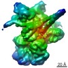
| Structure viewer | Molecule: MolmilJmol/JSmol |
|---|
- Downloads & links
-Download
| PDBx/mmCIF format | 5epi.cif.gz | 2.5 MB | Display | PDBx/mmCIF format |
|---|---|---|---|---|
| PDB format | pdb5epi.ent.gz | Display | PDB format | |
| PDBx/mmJSON format | 5epi.json.gz | Tree view | PDBx/mmJSON format | |
| Others |  Other downloads Other downloads |
-Validation report
| Arichive directory | https://data.pdbj.org/pub/pdb/validation_reports/ep/5epiftp://data.pdbj.org/pub/pdb/validation_reports/ep/5epi | HTTPS FTP |
|---|
-Related structure data
| Related structure data |  5fmlC  5fmmC  5fmqC  5fmzC  4wsaS S: Starting model for refinement C: citing same article ( |
|---|---|
| Similar structure data |


-Links
 PDBj
PDBj


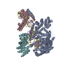
- Assembly
| Deposited unit | 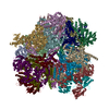
| ||||||||
|---|---|---|---|---|---|---|---|---|---|
| 1 | 
| ||||||||
| 2 | 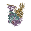
| ||||||||
| 3 | 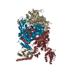
| ||||||||
| 4 | 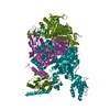
| ||||||||
| 5 | 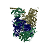
| ||||||||
| 6 | 
| ||||||||
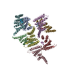
| Unit cell |
|
-Components
| #1: Protein | Mass: 85822.781 Da / Num. of mol.: 6 Source method: isolated from a genetically manipulated source Details: N-terminal extension, his-tag. C-terminal extension, linker and TEV site Source: (gene. exp.) Influenza B virus (B/Memphis/13/2003) / Gene: PA / Plasmid: PKL-PBAC / Cell line (production host): High Five / Production host:  Trichoplusia ni (cabbage looper) / References: UniProt: Q5V8Z9 Trichoplusia ni (cabbage looper) / References: UniProt: Q5V8Z9#2: Protein | Mass: 86207.016 Da / Num. of mol.: 6 / Fragment: PB1 SUBUNIT Source method: isolated from a genetically manipulated source Source: (gene. exp.) Influenza B virus (B/Memphis/13/2003) / Gene: PB1 / Plasmid: PKL-PBAC / Production host: Trichoplusia ni (cabbage looper) / References: UniProt: Q5V8Y6, RNA-directed RNA polymerase#3: Protein | Mass: 90858.133 Da / Num. of mol.: 6 Source method: isolated from a genetically manipulated source Source: (gene. exp.) Influenza B virus (B/Memphis/13/2003) / Gene: PB2 / Plasmid: PKL-PBAC / Production host: Trichoplusia ni (cabbage looper) / References: UniProt: Q5V8X3#4: RNA chain | Mass: 3921.464 Da / Num. of mol.: 6 / Fragment: FIRST 12 NUCLEOTIDES / Source method: obtained synthetically / Source: (synth.) Influenza B virus (B/Memphis/13/2003)Sequence details | N-TERMINAL EXTENSION, HIS-TAG AND LINKER. C-TERMINAL EXTENSION, LINKER AND TEV SITE. N-TERMINAL ...N-TERMINAL EXTENSION, HIS-TAG AND LINKER. C-TERMINAL EXTENSION, LINKER AND TEV SITE. N-TERMINAL EXTENSION, LINKER. C-TERMINAL EXTENSION, LINKER AND TEV SITE. N-TERMINAL EXTENSION, LINKER. C-TERMINAL EXTENSION, LINKER, STREP-TAG AND TEV SITE. | |
|---|
-Experimental details
-Experiment
| Experiment | Method: X-RAY DIFFRACTION / Number of used crystals: 1 |
|---|
- Sample preparation
| Crystal | Density Matthews: 4.2 Å3/Da / Density % sol: 70.7 % |
|---|---|
| Crystal grow | Temperature: 277 K / Method: vapor diffusion, sitting drop / pH: 5.6 Details: 100 MM TRI SODIUM CITRATE PH 5.6, 10 OR 100 MM NACL AND 12% MPD PH range: 5.6 |
-Data collection
| Diffraction | Mean temperature: 100 K |
|---|---|
| Diffraction source | Source: SYNCHROTRON / Site: ESRF / Beamline: ID29 / Wavelength: 1.07228 Å |
| Detector | Type: DECTRIS PILATUS 6M / Detector: PIXEL / Date: Apr 13, 2015 |
| Radiation | Protocol: SINGLE WAVELENGTH / Monochromatic (M) / Laue (L): M / Scattering type: x-ray |
| Radiation wavelength | Wavelength: 1.07228 Å / Relative weight: 1 |
| Reflection | Resolution: 4.1→50 Å / Num. obs: 195792 / % possible obs: 92.9 % / Observed criterion σ(I): 0 / Redundancy: 1.75 % / Rmerge(I) obs: 0.09 / Net I/σ(I): 5.49 |
| Reflection shell | Resolution: 4.1→4.3 Å / Redundancy: 1.77 % / Rmerge(I) obs: 0.63 / Mean I/σ(I) obs: 1.23 / % possible all: 94.9 |
- Processing
| Software |
| |||||||||||||||||||||||||||||||||||||||||||||||||||||||||||||||||||||||||||||||||||||||||||||||||||||||||||||||||||||||||||||||||||||||||||||||||||||||||||||||||||||||||||||||||||||||||||||||||||||||||||||||||||||||||
|---|---|---|---|---|---|---|---|---|---|---|---|---|---|---|---|---|---|---|---|---|---|---|---|---|---|---|---|---|---|---|---|---|---|---|---|---|---|---|---|---|---|---|---|---|---|---|---|---|---|---|---|---|---|---|---|---|---|---|---|---|---|---|---|---|---|---|---|---|---|---|---|---|---|---|---|---|---|---|---|---|---|---|---|---|---|---|---|---|---|---|---|---|---|---|---|---|---|---|---|---|---|---|---|---|---|---|---|---|---|---|---|---|---|---|---|---|---|---|---|---|---|---|---|---|---|---|---|---|---|---|---|---|---|---|---|---|---|---|---|---|---|---|---|---|---|---|---|---|---|---|---|---|---|---|---|---|---|---|---|---|---|---|---|---|---|---|---|---|---|---|---|---|---|---|---|---|---|---|---|---|---|---|---|---|---|---|---|---|---|---|---|---|---|---|---|---|---|---|---|---|---|---|---|---|---|---|---|---|---|---|---|---|---|---|---|---|---|---|
| Refinement | Method to determine structure: MOLECULAR REPLACEMENT Starting model: 4WSA Resolution: 4.1→49.945 Å / SU ML: 0.66 / Cross valid method: FREE R-VALUE / σ(F): 1.96 / Phase error: 33.77 / Stereochemistry target values: ML
| |||||||||||||||||||||||||||||||||||||||||||||||||||||||||||||||||||||||||||||||||||||||||||||||||||||||||||||||||||||||||||||||||||||||||||||||||||||||||||||||||||||||||||||||||||||||||||||||||||||||||||||||||||||||||
| Solvent computation | Shrinkage radii: 0.9 Å / VDW probe radii: 1.11 Å / Solvent model: FLAT BULK SOLVENT MODEL | |||||||||||||||||||||||||||||||||||||||||||||||||||||||||||||||||||||||||||||||||||||||||||||||||||||||||||||||||||||||||||||||||||||||||||||||||||||||||||||||||||||||||||||||||||||||||||||||||||||||||||||||||||||||||
| Refinement step | Cycle: LAST / Resolution: 4.1→49.945 Å
| |||||||||||||||||||||||||||||||||||||||||||||||||||||||||||||||||||||||||||||||||||||||||||||||||||||||||||||||||||||||||||||||||||||||||||||||||||||||||||||||||||||||||||||||||||||||||||||||||||||||||||||||||||||||||
| Refine LS restraints |
| |||||||||||||||||||||||||||||||||||||||||||||||||||||||||||||||||||||||||||||||||||||||||||||||||||||||||||||||||||||||||||||||||||||||||||||||||||||||||||||||||||||||||||||||||||||||||||||||||||||||||||||||||||||||||
| LS refinement shell |
|
