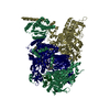
Journal: Mol Cell / Year: 2019 Title: Mechanism of DNA End Sensing and Processing by the Mre11-Rad50 Complex. Authors: Lisa Käshammer / Jan-Hinnerk Saathoff / Katja Lammens / Fabian Gut / Joseph Bartho / Aaron Alt / Brigitte Kessler / Karl-Peter Hopfner / Abstract: DNA double-strand breaks (DSBs) threaten genome stability throughout life and are linked to tumorigenesis in humans. To initiate DSB repair by end joining or homologous recombination, the Mre11- ...DNA double-strand breaks (DSBs) threaten genome stability throughout life and are linked to tumorigenesis in humans. To initiate DSB repair by end joining or homologous recombination, the Mre11-nuclease Rad50-ATPase complex detects and processes diverse and obstructed DNA ends, but a structural mechanism is still lacking. Here we report cryo-EM structures of the E. coli Mre11-Rad50 homolog SbcCD in resting and DNA-bound cutting states. In the resting state, Mre11's nuclease is blocked by ATP-Rad50, and the Rad50 coiled coils appear flexible. Upon DNA binding, the two coiled coils zip up into a rod and, together with the Rad50 nucleotide-binding domains, form a clamp around dsDNA. Mre11 moves to the side of Rad50, binds the DNA end, and assembles a DNA cutting channel for the nuclease reactions. The structures reveal how Mre11-Rad50 can detect and process diverse DNA ends and uncover a clamping and gating function for the coiled coils.
History
Deposition
Jul 8, 2019
-
Header (metadata) release
Sep 4, 2019
-
Map release
Sep 4, 2019
-
Update
Nov 20, 2019
-
Current status
Nov 20, 2019
Processing site: PDBe / Status: Released
-
Structure visualization
Movie
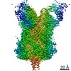
Surface view with section colored by density value
Entire : Heterotetrameric complex of full-length Mre11-Rad50 (SbcCD) in co...
Entire
Name: Heterotetrameric complex of full-length Mre11-Rad50 (SbcCD) in complex with ADP and dsDNA
Components
Complex: Heterotetrameric complex of full-length Mre11-Rad50 (SbcCD) in complex with ADP and dsDNA
-
Supramolecule #1: Heterotetrameric complex of full-length Mre11-Rad50 (SbcCD) in co...
Supramolecule
Name: Heterotetrameric complex of full-length Mre11-Rad50 (SbcCD) in complex with ADP and dsDNA type: complex / ID: 1 / Parent: 0 / Macromolecule list: #1-#4
Source (natural)
Organism: Escherichia coli (E. coli)
Recombinant expression
Organism: Escherichia coli BL21(DE3) (bacteria)
-
Experimental details
-
Structure determination
Method
cryo EM
Processing
single particle reconstruction
Aggregation state
particle
-
Sample preparation
Buffer
pH: 7.5
Vitrification
Cryogen name: ETHANE / Chamber humidity: 95 % / Chamber temperature: 277 K / Instrument: FEI VITROBOT MARK IV
-
Electron microscopy
Microscope
FEI TITAN KRIOS
Image recording
Film or detector model: GATAN K2 SUMMIT (4k x 4k) / Detector mode: COUNTING / Number grids imaged: 2 / Number real images: 12811 / Average electron dose: 73.6 e/Å2
Electron beam
Acceleration voltage: 300 kV / Electron source: FIELD EMISSION GUN
Electron optics
Illumination mode: FLOOD BEAM / Imaging mode: BRIGHT FIELD
Experimental equipment
Model: Titan Krios / Image courtesy: FEI Company
-
Image processing
Startup model
Type of model: NONE Details: The initial model was calculated de novo using cryoSPARC.
Final reconstruction
Resolution.type: BY AUTHOR / Resolution: 4.3 Å / Resolution method: FSC 0.143 CUT-OFF / Number images used: 152271
Initial angle assignment
Type: MAXIMUM LIKELIHOOD
Final angle assignment
Type: MAXIMUM LIKELIHOOD
+
About Yorodumi
-
News
-
Feb 9, 2022. New format data for meta-information of EMDB entries
New format data for meta-information of EMDB entries
Version 3 of the EMDB header file is now the official format.
The previous official version 1.9 will be removed from the archive.
In the structure databanks used in Yorodumi, some data are registered as the other names, "COVID-19 virus" and "2019-nCoV". Here are the details of the virus and the list of structure data.
Jan 31, 2019. EMDB accession codes are about to change! (news from PDBe EMDB page)
EMDB accession codes are about to change! (news from PDBe EMDB page)
The allocation of 4 digits for EMDB accession codes will soon come to an end. Whilst these codes will remain in use, new EMDB accession codes will include an additional digit and will expand incrementally as the available range of codes is exhausted. The current 4-digit format prefixed with “EMD-” (i.e. EMD-XXXX) will advance to a 5-digit format (i.e. EMD-XXXXX), and so on. It is currently estimated that the 4-digit codes will be depleted around Spring 2019, at which point the 5-digit format will come into force.
The EM Navigator/Yorodumi systems omit the EMD- prefix.
Related info.:Q: What is EMD? / ID/Accession-code notation in Yorodumi/EM Navigator
Yorodumi is a browser for structure data from EMDB, PDB, SASBDB, etc.
This page is also the successor to EM Navigator detail page, and also detail information page/front-end page for Omokage search.
The word "yorodu" (or yorozu) is an old Japanese word meaning "ten thousand". "mi" (miru) is to see.
Related info.:EMDB / PDB / SASBDB / Comparison of 3 databanks / Yorodumi Search / Aug 31, 2016. New EM Navigator & Yorodumi / Yorodumi Papers / Jmol/JSmol / Function and homology information / Changes in new EM Navigator and Yorodumi
 Movie
Movie Controller

 Yorodumi
 Open data
 Basic information
 Map data
 Sample
 Function and homology information
 Authors
 Germany, 2 items
  Citation
 Structure visualization
 Downloads & links
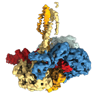 emd_10114.png
 http://ftp.pdbj.org/pub/emdb/structures/EMD-10114


 Z (Sec.)
 Y (Row.)
 X (Col.)


 Sample components
 Processing
 Electron microscopy
 FIELD EMISSION GUN
