 Movie
Movie Controller

[English] 日本語
 Yorodumi
- PDB-4wsa: Crystal structure of Influenza B polymerase bound to the vRNA pro... -
+ Open data

- Basic information
| Entry | Database: PDB / ID: 4wsa | ||||||
|---|---|---|---|---|---|---|---|
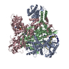
| Title | Crystal structure of Influenza B polymerase bound to the vRNA promoter (FluB1 form) | ||||||
 Components Components |
| ||||||
 Keywords Keywords | TRANSFERASE/RNA / TRANSFERASE-RNA COMPLEX | ||||||
| Function / homology |  Function and homology information Function and homology informationcap snatching / viral transcription / symbiont-mediated suppression of host mRNA transcription via inhibition of RNA polymerase II activity / host cell mitochondrion / 7-methylguanosine mRNA capping / virion component / endonuclease activity / Hydrolases; Acting on ester bonds / host cell cytoplasm / symbiont-mediated suppression of host gene expression ...cap snatching / viral transcription / symbiont-mediated suppression of host mRNA transcription via inhibition of RNA polymerase II activity / host cell mitochondrion / 7-methylguanosine mRNA capping / virion component / endonuclease activity / Hydrolases; Acting on ester bonds / host cell cytoplasm / symbiont-mediated suppression of host gene expression / viral translational frameshifting / RNA-directed RNA polymerase / nucleotide binding / viral RNA genome replication / hydrolase activity / RNA-directed RNA polymerase activity / host cell nucleus / DNA-templated transcription / RNA binding / metal ion binding Similarity search - Function | ||||||
| Biological species |  Influenza B virus Influenza B virus | ||||||
| Method |  X-RAY DIFFRACTION / SYNCHROTRON / SIRAS / Resolution: 3.4 Å X-RAY DIFFRACTION / SYNCHROTRON / SIRAS / Resolution: 3.4 Å | ||||||
 Authors Authors | Reich, S. / Guilligay, D. / Pflug, A. / Cusack, S. | ||||||
 Citation Citation | Journal: Nature / Year: 2014 Title: Structural insight into cap-snatching and RNA synthesis by influenza polymerase. Authors: Reich, S. / Guilligay, D. / Pflug, A. / Malet, H. / Berger, I. / Crepin, T. / Hart, D. / Lunardi, T. / Nanao, M. / Ruigrok, R.W. / Cusack, S. | ||||||
| History |
|
- Structure visualization
| Structure viewer | Molecule: MolmilJmol/JSmol |
|---|
- Downloads & links
-Download
| PDBx/mmCIF format | 4wsa.cif.gz | 467.2 KB | Display | PDBx/mmCIF format |
|---|---|---|---|---|
| PDB format | pdb4wsa.ent.gz | 368.9 KB | Display | PDB format |
| PDBx/mmJSON format | 4wsa.json.gz | Tree view | PDBx/mmJSON format | |
| Others |  Other downloads Other downloads |
-Validation report
| Arichive directory | https://data.pdbj.org/pub/pdb/validation_reports/ws/4wsaftp://data.pdbj.org/pub/pdb/validation_reports/ws/4wsa | HTTPS FTP |
|---|
-Related structure data


-Links
 PDBj
PDBj


- Assembly
| Deposited unit | 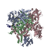
| ||||||||
|---|---|---|---|---|---|---|---|---|---|
| 1 |
| ||||||||
| Unit cell |
|
-Components
| #1: RNA chain | Mass: 5584.283 Da / Num. of mol.: 1 / Source method: obtained synthetically / Source: (synth.) Influenza B virus |
|---|---|
| #2: RNA chain | Mass: 4557.820 Da / Num. of mol.: 1 / Source method: obtained synthetically / Source: (synth.) Influenza B virus |
| #3: Protein | Mass: 85822.781 Da / Num. of mol.: 1 Source method: isolated from a genetically manipulated source Details: N-terminal extension: his-tag and linker C-terminal extension: linker and TEV site Source: (gene. exp.) Influenza B virus / Strain: B/Memphis/13/03 / Gene: PA / Plasmid: pKL-PBac / Cell line (production host): High Five / Production host:  Trichoplusia ni (cabbage looper) / References: UniProt: Q5V8Z9 Trichoplusia ni (cabbage looper) / References: UniProt: Q5V8Z9 |
| #4: Protein | Mass: 86207.016 Da / Num. of mol.: 1 / Mutation: Trichoplusia ni Source method: isolated from a genetically manipulated source Details: N-terminal extension: linker C-terminal extension: linker and TEV site Source: (gene. exp.) Influenza B virus / Strain: B/Memphis/13/03 / Gene: PB1 / Plasmid: pKL-PBac / Cell line (production host): High Five / Production host: Trichoplusia ni (cabbage looper) / References: UniProt: Q5V8Y6, RNA-directed RNA polymerase |
| #5: Protein | Mass: 90844.109 Da / Num. of mol.: 1 Source method: isolated from a genetically manipulated source Details: N-terminal extension: linker C-terminal extension: Strep-tag and TEV site Source: (gene. exp.) Influenza B virus / Strain: B/Memphis/13/03 / Gene: PB2 / Plasmid: pKL-PBac / Cell line (production host): High Five / Production host: Trichoplusia ni (cabbage looper) / References: UniProt: Q5V8X3 |
-Experimental details
-Experiment
| Experiment | Method: X-RAY DIFFRACTION |
|---|
- Sample preparation
| Crystal | Density Matthews: 5.33 Å3/Da / Density % sol: 75 % / Description: bipyramids |
|---|---|
| Crystal grow | Temperature: 281 K / Method: vapor diffusion, sitting drop / pH: 9 Details: FluB polymerase was concentrated to 9 mg per ml (37 microM) in a buffer containing 500 mM NaCl, 50 mM Hepes pH 7.5, 5% glycerol and 2mM Tris(2-carboxyethyl)phosphine (TCEP), and mixed with ...Details: FluB polymerase was concentrated to 9 mg per ml (37 microM) in a buffer containing 500 mM NaCl, 50 mM Hepes pH 7.5, 5% glycerol and 2mM Tris(2-carboxyethyl)phosphine (TCEP), and mixed with 40 microM vRNA for crystallization in hanging drops at 4 C. A trigonal crystal form (FluB1) was obtained by mixing polymerase with nucleotides 5-18 of the 3 prime end and 1-14 of the 5 prime end of the vRNA in a condition containing 0.1 M bicine pH 9.0, 10% MPD. |
-Data collection
| Diffraction | Mean temperature: 100 K |
|---|---|
| Diffraction source | Source: SYNCHROTRON / Site: ESRF  / Beamline: ID23-1 / Wavelength: 0.973 Å / Beamline: ID23-1 / Wavelength: 0.973 Å |
| Detector | Type: DECTRIS PILATUS 6M-F / Detector: PIXEL / Date: Apr 4, 2014 |
| Radiation | Protocol: SINGLE WAVELENGTH / Monochromatic (M) / Laue (L): M / Scattering type: x-ray |
| Radiation wavelength | Wavelength: 0.973 Å / Relative weight: 1 |
| Reflection | Resolution: 3.4→50 Å / Num. obs: 79266 / % possible obs: 98.5 % / Observed criterion σ(F): 0 / Observed criterion σ(I): 0 / Redundancy: 11.8 % / Rmerge(I) obs: 0.121 / Net I/σ(I): 12.6 |
| Reflection shell | Resolution: 3.4→3.52 Å / Redundancy: 5.8 % / Rmerge(I) obs: 1.3 / Mean I/σ(I) obs: 1.5 / % possible all: 89.9 |
- Processing
| Software |
| ||||||||||||||||||||||||||||||||||||||||||||||||||||||||||||||||||||||||||||||||||||||||||||||||||||||||||||||||||||||||||||||||||||||||||||||||||||||||||||||||||||||||||||||||||||||||||||||||||||
|---|---|---|---|---|---|---|---|---|---|---|---|---|---|---|---|---|---|---|---|---|---|---|---|---|---|---|---|---|---|---|---|---|---|---|---|---|---|---|---|---|---|---|---|---|---|---|---|---|---|---|---|---|---|---|---|---|---|---|---|---|---|---|---|---|---|---|---|---|---|---|---|---|---|---|---|---|---|---|---|---|---|---|---|---|---|---|---|---|---|---|---|---|---|---|---|---|---|---|---|---|---|---|---|---|---|---|---|---|---|---|---|---|---|---|---|---|---|---|---|---|---|---|---|---|---|---|---|---|---|---|---|---|---|---|---|---|---|---|---|---|---|---|---|---|---|---|---|---|---|---|---|---|---|---|---|---|---|---|---|---|---|---|---|---|---|---|---|---|---|---|---|---|---|---|---|---|---|---|---|---|---|---|---|---|---|---|---|---|---|---|---|---|---|---|---|---|---|
| Refinement | Method to determine structure: SIRAS / Resolution: 3.4→48.978 Å / SU ML: 0.48 / Cross valid method: FREE R-VALUE / σ(F): 1.36 / Phase error: 32.94 / Stereochemistry target values: ML
| ||||||||||||||||||||||||||||||||||||||||||||||||||||||||||||||||||||||||||||||||||||||||||||||||||||||||||||||||||||||||||||||||||||||||||||||||||||||||||||||||||||||||||||||||||||||||||||||||||||
| Solvent computation | Shrinkage radii: 0.9 Å / VDW probe radii: 1.11 Å / Solvent model: FLAT BULK SOLVENT MODEL | ||||||||||||||||||||||||||||||||||||||||||||||||||||||||||||||||||||||||||||||||||||||||||||||||||||||||||||||||||||||||||||||||||||||||||||||||||||||||||||||||||||||||||||||||||||||||||||||||||||
| Refinement step | Cycle: 1 / Resolution: 3.4→48.978 Å
| ||||||||||||||||||||||||||||||||||||||||||||||||||||||||||||||||||||||||||||||||||||||||||||||||||||||||||||||||||||||||||||||||||||||||||||||||||||||||||||||||||||||||||||||||||||||||||||||||||||
| Refine LS restraints |
| ||||||||||||||||||||||||||||||||||||||||||||||||||||||||||||||||||||||||||||||||||||||||||||||||||||||||||||||||||||||||||||||||||||||||||||||||||||||||||||||||||||||||||||||||||||||||||||||||||||
| LS refinement shell |
|
