 Movie
Movie Controller

[English] 日本語
 Yorodumi
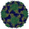
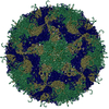
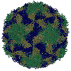
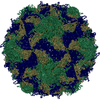
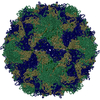
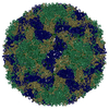
- PDB-5ddj: Crystal structure of recombinant foot-and-mouth-disease virus O1M... -
+ Open data

- Basic information
| Entry | Database: PDB / ID: 5ddj | ||||||
|---|---|---|---|---|---|---|---|
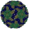
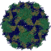
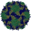
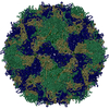
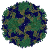
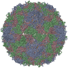
| Title | Crystal structure of recombinant foot-and-mouth-disease virus O1M-S2093Y empty capsid | ||||||
 Components Components |
| ||||||
 Keywords Keywords | VIRUS / foot and mouth disease virus / picornavirus / aphthovirus / vaccine | ||||||
| Function / homology |  Function and homology information Function and homology informationsymbiont-mediated perturbation of host chromatin organization / T=pseudo3 icosahedral viral capsid / host cell cytoplasmic vesicle membrane / ribonucleoside triphosphate phosphatase activity / channel activity / monoatomic ion transmembrane transport / clathrin-dependent endocytosis of virus by host cell / RNA helicase activity / viral protein processing / host cell endoplasmic reticulum membrane ...symbiont-mediated perturbation of host chromatin organization / T=pseudo3 icosahedral viral capsid / host cell cytoplasmic vesicle membrane / ribonucleoside triphosphate phosphatase activity / channel activity / monoatomic ion transmembrane transport / clathrin-dependent endocytosis of virus by host cell / RNA helicase activity / viral protein processing / host cell endoplasmic reticulum membrane / cysteine-type endopeptidase activity / viral RNA genome replication / RNA-directed RNA polymerase activity / virion attachment to host cell / host cell nucleus / DNA-templated transcription / structural molecule activity / proteolysis / RNA binding / ATP binding Similarity search - Function | ||||||
| Biological species |  Foot-and-mouth disease virus - type O Foot-and-mouth disease virus - type O | ||||||
| Method |  X-RAY DIFFRACTION / SYNCHROTRON / MOLECULAR REPLACEMENT / Resolution: 3.5 Å X-RAY DIFFRACTION / SYNCHROTRON / MOLECULAR REPLACEMENT / Resolution: 3.5 Å | ||||||
 Authors Authors | Kotecha, A. / Seago, J. / Scott, K. / Burman, A. / Loureiro, S. / Ren, J. / Porta, C. / Ginn, H.M. / Jackson, T. / Perez-Martin, E. ...Kotecha, A. / Seago, J. / Scott, K. / Burman, A. / Loureiro, S. / Ren, J. / Porta, C. / Ginn, H.M. / Jackson, T. / Perez-Martin, E. / Siebert, C.A. / Paul, G. / Huiskonen, J.T. / Jones, I.M. / Esnouf, R.M. / Fry, E.E. / Maree, F.F. / Charleston, B. / Stuart, D.I. | ||||||
| Funding support |  United Kingdom, 1items United Kingdom, 1items
| ||||||
 Citation Citation | Journal: Nat Struct Mol Biol / Year: 2015 Title: Structure-based energetics of protein interfaces guides foot-and-mouth disease virus vaccine design. Authors: Abhay Kotecha / Julian Seago / Katherine Scott / Alison Burman / Silvia Loureiro / Jingshan Ren / Claudine Porta / Helen M Ginn / Terry Jackson / Eva Perez-Martin / C Alistair Siebert / ...Authors: Abhay Kotecha / Julian Seago / Katherine Scott / Alison Burman / Silvia Loureiro / Jingshan Ren / Claudine Porta / Helen M Ginn / Terry Jackson / Eva Perez-Martin / C Alistair Siebert / Guntram Paul / Juha T Huiskonen / Ian M Jones / Robert M Esnouf / Elizabeth E Fry / Francois F Maree / Bryan Charleston / David I Stuart /   Abstract: Virus capsids are primed for disassembly, yet capsid integrity is key to generating a protective immune response. Foot-and-mouth disease virus (FMDV) capsids comprise identical pentameric protein ...Virus capsids are primed for disassembly, yet capsid integrity is key to generating a protective immune response. Foot-and-mouth disease virus (FMDV) capsids comprise identical pentameric protein subunits held together by tenuous noncovalent interactions and are often unstable. Chemically inactivated or recombinant empty capsids, which could form the basis of future vaccines, are even less stable than live virus. Here we devised a computational method to assess the relative stability of protein-protein interfaces and used it to design improved candidate vaccines for two poorly stable, but globally important, serotypes of FMDV: O and SAT2. We used a restrained molecular dynamics strategy to rank mutations predicted to strengthen the pentamer interfaces and applied the results to produce stabilized capsids. Structural analyses and stability assays confirmed the predictions, and vaccinated animals generated improved neutralizing-antibody responses to stabilized particles compared to parental viruses and wild-type capsids. | ||||||
| History |
|
- Structure visualization
| Structure viewer | Molecule: MolmilJmol/JSmol |
|---|
- Downloads & links
-Download
| PDBx/mmCIF format | 5ddj.cif.gz | 145.5 KB | Display | PDBx/mmCIF format |
|---|---|---|---|---|
| PDB format | pdb5ddj.ent.gz | 111.7 KB | Display | PDB format |
| PDBx/mmJSON format | 5ddj.json.gz | Tree view | PDBx/mmJSON format | |
| Others |  Other downloads Other downloads |
-Validation report
| Arichive directory | https://data.pdbj.org/pub/pdb/validation_reports/dd/5ddjftp://data.pdbj.org/pub/pdb/validation_reports/dd/5ddj | HTTPS FTP |
|---|
-Related structure data
| Related structure data | 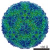 3129C 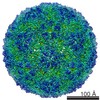 3130C 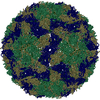 5ac9C  5acaC  5d8aC  1fodS S: Starting model for refinement C: citing same article ( |
|---|---|
| Similar structure data |


-Links
 PDBj
PDBj


- Assembly
| Deposited unit | 
| ||||||||||||||||||
|---|---|---|---|---|---|---|---|---|---|---|---|---|---|---|---|---|---|---|---|
| 1 | x 60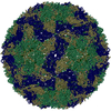
| ||||||||||||||||||
| 2 |
| ||||||||||||||||||
| 3 | x 5
| ||||||||||||||||||
| 4 | x 6
| ||||||||||||||||||
| 5 | 
| ||||||||||||||||||
| Unit cell |
| ||||||||||||||||||
| Symmetry | Point symmetry: (Schoenflies symbol: I (icosahedral)) | ||||||||||||||||||
| Noncrystallographic symmetry (NCS) | NCS oper:
|
-Components
| #1: Protein | Mass: 23207.336 Da / Num. of mol.: 1 Source method: isolated from a genetically manipulated source Source: (gene. exp.) Foot-and-mouth disease virus - type O / Cell line (production host): BHK-21, Clone 13 / Production host:  MESOCRICETUS AURATUS (golden hamster) / Strain (production host): BHK-21 / References: UniProt: Q6PMW3 MESOCRICETUS AURATUS (golden hamster) / Strain (production host): BHK-21 / References: UniProt: Q6PMW3 |
|---|---|
| #2: Protein | Mass: 24417.510 Da / Num. of mol.: 1 / Mutation: S93Y Source method: isolated from a genetically manipulated source Source: (gene. exp.) Foot-and-mouth disease virus - type O / Cell line (production host): BHK-21, Clone 13 / Production host: MESOCRICETUS AURATUS (golden hamster) / Strain (production host): BHK-21 / References: UniProt: Q6PMW3 |
| #3: Protein | Mass: 23905.826 Da / Num. of mol.: 1 Source method: isolated from a genetically manipulated source Source: (gene. exp.) Foot-and-mouth disease virus - type O / Cell line (production host): BHK-21, Clone 13 / Production host: MESOCRICETUS AURATUS (golden hamster) / Strain (production host): BHK-21 / References: UniProt: Q6PMW3 |
| #4: Protein | Mass: 8766.075 Da / Num. of mol.: 1 Source method: isolated from a genetically manipulated source Source: (gene. exp.) Foot-and-mouth disease virus - type O / Cell line (production host): BHK-21, Clone 13 / Production host: MESOCRICETUS AURATUS (golden hamster) / Strain (production host): BHK-21 / References: UniProt: Q6PMW3 |
-Experimental details
-Experiment
| Experiment | Method: X-RAY DIFFRACTION / Number of used crystals: 1 |
|---|
- Sample preparation
| Crystal grow | Temperature: 298 K / Method: vapor diffusion, sitting drop / pH: 7 Details: 1.5 M ammonium sulphate, 100 mM bis-Tris Propane, pH 7.0 PH range: 7 |
|---|
-Data collection
| Diffraction | Mean temperature: 298 K | ||||||||||||||||||||||||||||||||||||||||||||||||||||||||||||||||||
|---|---|---|---|---|---|---|---|---|---|---|---|---|---|---|---|---|---|---|---|---|---|---|---|---|---|---|---|---|---|---|---|---|---|---|---|---|---|---|---|---|---|---|---|---|---|---|---|---|---|---|---|---|---|---|---|---|---|---|---|---|---|---|---|---|---|---|---|
| Diffraction source | Source: SYNCHROTRON / Site: Diamond / Beamline: I24 / Wavelength: 0.9778 Å | ||||||||||||||||||||||||||||||||||||||||||||||||||||||||||||||||||
| Detector | Type: PSI PILATUS 6M / Detector: PIXEL / Date: May 10, 2012 | ||||||||||||||||||||||||||||||||||||||||||||||||||||||||||||||||||
| Radiation | Protocol: SINGLE WAVELENGTH / Monochromatic (M) / Laue (L): M / Scattering type: x-ray | ||||||||||||||||||||||||||||||||||||||||||||||||||||||||||||||||||
| Radiation wavelength | Wavelength: 0.9778 Å / Relative weight: 1 | ||||||||||||||||||||||||||||||||||||||||||||||||||||||||||||||||||
| Reflection | Redundancy: 1.5 % / Number: 67562 / Rmerge(I) obs: 0.427 / Χ2: 0.93 / D res high: 3.5 Å / D res low: 50 Å / Num. obs: 45271 / % possible obs: 53.2 | ||||||||||||||||||||||||||||||||||||||||||||||||||||||||||||||||||
| Diffraction reflection shell |
| ||||||||||||||||||||||||||||||||||||||||||||||||||||||||||||||||||
| Reflection | Resolution: 3.5→50 Å / Num. obs: 45271 / % possible obs: 53.2 % / Redundancy: 1.5 % / Rmerge(I) obs: 0.427 / Χ2: 0.928 / Net I/av σ(I): 1.547 / Net I/σ(I): 1.7 / Num. measured all: 67562 | ||||||||||||||||||||||||||||||||||||||||||||||||||||||||||||||||||
| Reflection shell | Diffraction-ID: 1 / Rejects: _
|
- Processing
| Software |
| ||||||||||||||||||||||||||||||||||||||||||||||||||||||||||||||||||
|---|---|---|---|---|---|---|---|---|---|---|---|---|---|---|---|---|---|---|---|---|---|---|---|---|---|---|---|---|---|---|---|---|---|---|---|---|---|---|---|---|---|---|---|---|---|---|---|---|---|---|---|---|---|---|---|---|---|---|---|---|---|---|---|---|---|---|---|
| Refinement | Method to determine structure: MOLECULAR REPLACEMENT Starting model: 1FOD Resolution: 3.5→49.664 Å / Cross valid method: NONE / σ(F): 0
| ||||||||||||||||||||||||||||||||||||||||||||||||||||||||||||||||||
| Solvent computation | Bsol: 75.2315 Å2 | ||||||||||||||||||||||||||||||||||||||||||||||||||||||||||||||||||
| Displacement parameters | Biso max: 135.49 Å2 / Biso mean: 47.2234 Å2 / Biso min: 3 Å2
| ||||||||||||||||||||||||||||||||||||||||||||||||||||||||||||||||||
| Refinement step | Cycle: final / Resolution: 3.5→49.664 Å
| ||||||||||||||||||||||||||||||||||||||||||||||||||||||||||||||||||
| Refine LS restraints |
| ||||||||||||||||||||||||||||||||||||||||||||||||||||||||||||||||||
| LS refinement shell |
| ||||||||||||||||||||||||||||||||||||||||||||||||||||||||||||||||||
| Xplor file |
|
