[English] 日本語
Yorodumi
- PDB-4ypn: Crystal structure of a LonA fragment containing the 3-helix bundl... -

+
Open data


ID or keywords:

Loading...

-
Basic information

Entry
Database: PDB / ID: 4ypn
TitleCrystal structure of a LonA fragment containing the 3-helix bundle and the AAA-alpha/beta domain
ComponentsLon proteaseLon protease family
KeywordsHYDROLASE / AAA+ domain / Helix-bundle / disordered
Function / homology
Function and homology information


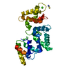
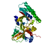
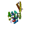
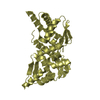
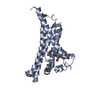
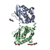
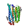
endopeptidase La / ATP-dependent peptidase activity / protein quality control for misfolded or incompletely synthesized proteins / cellular response to heat / sequence-specific DNA binding / serine-type endopeptidase activity / ATP hydrolysis activity / ATP binding / identical protein binding / cytoplasm
Similarity search - Function
Lon protease, bacterial / Lon protease, bacterial/eukaryotic-type / Peptidase S16, active site / ATP-dependent serine proteases, lon family, serine active site. / Lon proteolytic domain profile. / Peptidase S16, Lon proteolytic domain / Lon protease / Lon protease (S16) C-terminal proteolytic domain / Lon protease, N-terminal domain superfamily / Lon N-terminal domain profile. ...Lon protease, bacterial / Lon protease, bacterial/eukaryotic-type / Peptidase S16, active site / ATP-dependent serine proteases, lon family, serine active site. / Lon proteolytic domain profile. / Peptidase S16, Lon proteolytic domain / Lon protease / Lon protease (S16) C-terminal proteolytic domain / Lon protease, N-terminal domain superfamily / Lon N-terminal domain profile. / Lon protease, N-terminal domain / ATP-dependent protease La (LON) substrate-binding domain / Found in ATP-dependent protease La (LON) / PUA-like superfamily / ATPase family associated with various cellular activities (AAA) / ATPase, AAA-type, core / Ribosomal protein S5 domain 2-type fold, subgroup / Ribosomal protein S5 domain 2-type fold / ATPases associated with a variety of cellular activities / AAA+ ATPase domain / P-loop containing nucleoside triphosphate hydrolase
Similarity search - Domain/homology
Biological speciesMeiothermus taiwanensis (bacteria)
MethodX-RAY DIFFRACTION / SYNCHROTRON / MOLECULAR REPLACEMENT / Resolution: 2.07 Å
AuthorsChang, C.-I. / Lin, C.-C.
Funding support Taiwan, 2items
OrganizationGrant numberCountry
National Science Council of TaiwanNSC102-2320-B-001-016 Taiwan
Academia Sinica Taiwan
CitationJournal: Structure / Year: 2016
Title: Structural Insights into the Allosteric Operation of the Lon AAA+ Protease
Authors: Lin, C.-C. / Su, S.-C. / Su, M.-Y. / Liang, P.-H. / Feng, C.-C. / Wu, S.-H. / Chang, C.-I.
History
DepositionMar 13, 2015Deposition site: RCSB / Processing site: PDBJ
Revision 1.0Mar 16, 2016Provider: repository / Type: Initial release
Revision 1.1Apr 20, 2016Group: Database references
Revision 1.2May 18, 2016Group: Database references
Revision 1.3Feb 5, 2020Group: Data collection / Database references / Derived calculations
Category: citation / diffrn_source / pdbx_struct_oper_list
Item: _citation.journal_id_CSD / _diffrn_source.pdbx_synchrotron_site / _pdbx_struct_oper_list.symmetry_operation
Revision 1.4Nov 8, 2023Group: Data collection / Database references / Refinement description
Category: chem_comp_atom / chem_comp_bond ...chem_comp_atom / chem_comp_bond / database_2 / pdbx_initial_refinement_model
Item: _database_2.pdbx_DOI / _database_2.pdbx_database_accession

-
Structure visualization

Structure viewerMolecule:
MolmilJmol/JSmol

Downloads & links

-
Assembly

Deposited unit
A: Lon protease


Theoretical massNumber of molelcules
Total (without water)33,5711
Polymers33,5711
Non-polymers00
Water1,53185
1


  • Idetical with deposited unit
  • defined by author
TypeNameSymmetry operationNumber
identity operation1_555x,y,z1
Buried area0 Å2
ΔGint0 kcal/mol
Surface area12810 Å2
Unit cell
Length a, b, c (Å)59.203, 63.892, 69.298
Angle α, β, γ (deg.)90.00, 90.00, 90.00
Int Tables number18
Space group name H-MP21221

-
Components

#1: Protein Lon protease / Lon protease family / ATP-dependent protease La


Mass: 33571.477 Da / Num. of mol.: 1
Fragment: 3-Helix-bundle and alpha/beta subdomain, UNP residues 207-492
Source method: isolated from a genetically manipulated source
Source: (gene. exp.) Meiothermus taiwanensis (bacteria) / Gene: lonA1, lon / Production host: Escherichia coli BL21(DE3) (bacteria) / Strain (production host): BL21(DE3) / References: UniProt: A0A059VAZ3, endopeptidase La
#2: Water ChemComp-HOH / water / Water


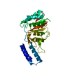
Mass: 18.015 Da / Num. of mol.: 85 / Source method: isolated from a natural source / Formula: H2O

-
Experimental details

-
Experiment

ExperimentMethod: X-RAY DIFFRACTION / Number of used crystals: 1

-
Sample preparation

CrystalDensity Matthews: 1.96 Å3/Da / Density % sol: 37.37 %
Crystal growTemperature: 295 K / Method: evaporation / pH: 5.5 / Details: 0.1 M sodium citrate pH 5.5, 25 % PEG 3350

-
Data collection

DiffractionMean temperature: 200 K
Diffraction sourceSource: SYNCHROTRON / Site: Photon Factory / Beamline: BL-1A / Wavelength: 1.1 Å
DetectorType: DECTRIS PILATUS 2M-F / Detector: PIXEL / Date: Jan 27, 2013
RadiationProtocol: SINGLE WAVELENGTH / Monochromatic (M) / Laue (L): M / Scattering type: x-ray
Radiation wavelengthWavelength: 1.1 Å / Relative weight: 1
ReflectionResolution: 2.07→50 Å / Num. all: 16502 / Num. obs: 15628 / % possible obs: 99.8 % / Observed criterion σ(I): 2 / Redundancy: 6.1 % / Rmerge(I) obs: 0.086 / Net I/σ(I): 19.49
Reflection shellResolution: 2.07→2.14 Å / Redundancy: 6.2 % / Rmerge(I) obs: 0.575 / Mean I/σ(I) obs: 4.62 / % possible all: 100

-
Processing

Software
NameVersionClassification
REFMAC5.8.0073refinement
HKL-2000data processing
HKL-2000data scaling
RefinementMethod to determine structure: MOLECULAR REPLACEMENT
Starting model: 3M6A
Resolution: 2.07→30 Å / Cor.coef. Fo:Fc: 0.958 / Cor.coef. Fo:Fc free: 0.931 / SU B: 10.321 / SU ML: 0.134 / Cross valid method: THROUGHOUT / ESU R: 0.207 / ESU R Free: 0.182 / Stereochemistry target values: MAXIMUM LIKELIHOOD / Details: HYDROGENS HAVE BEEN ADDED IN THE RIDING POSITIONS
RfactorNum. reflection% reflectionSelection details
Rfree0.23657 833 5.1 %RANDOM
Rwork0.18488 ---
obs0.18747 15627 99.19 %-
Solvent computationIon probe radii: 0.8 Å / Shrinkage radii: 0.8 Å / VDW probe radii: 1.2 Å / Solvent model: MASK
Displacement parametersBiso mean: 37.758 Å2
Baniso -1Baniso -2Baniso -3
1-0.39 Å20 Å20 Å2
2---0.42 Å20 Å2
3---0.03 Å2
Refinement stepCycle: LAST / Resolution: 2.07→30 Å
ProteinNucleic acidLigandSolventTotal
Num. atoms1985 0 0 85 2070
Refine LS restraints
Refine-IDTypeDev idealDev ideal targetNumber
X-RAY DIFFRACTIONr_bond_refined_d0.0180.0192021
X-RAY DIFFRACTIONr_bond_other_d0.0050.022005
X-RAY DIFFRACTIONr_angle_refined_deg1.8921.9852736
X-RAY DIFFRACTIONr_angle_other_deg0.94734619
X-RAY DIFFRACTIONr_dihedral_angle_1_deg6.9815249
X-RAY DIFFRACTIONr_dihedral_angle_2_deg36.41323.73691
X-RAY DIFFRACTIONr_dihedral_angle_3_deg15.62715375
X-RAY DIFFRACTIONr_dihedral_angle_4_deg22.7011519
X-RAY DIFFRACTIONr_chiral_restr0.1320.2313
X-RAY DIFFRACTIONr_gen_planes_refined0.0080.0212233
X-RAY DIFFRACTIONr_gen_planes_other0.0010.02426
X-RAY DIFFRACTIONr_nbd_refined
X-RAY DIFFRACTIONr_nbd_other
X-RAY DIFFRACTIONr_nbtor_refined
X-RAY DIFFRACTIONr_nbtor_other
X-RAY DIFFRACTIONr_xyhbond_nbd_refined
X-RAY DIFFRACTIONr_xyhbond_nbd_other
X-RAY DIFFRACTIONr_metal_ion_refined
X-RAY DIFFRACTIONr_metal_ion_other
X-RAY DIFFRACTIONr_symmetry_vdw_refined
X-RAY DIFFRACTIONr_symmetry_vdw_other
X-RAY DIFFRACTIONr_symmetry_hbond_refined
X-RAY DIFFRACTIONr_symmetry_hbond_other
X-RAY DIFFRACTIONr_symmetry_metal_ion_refined
X-RAY DIFFRACTIONr_symmetry_metal_ion_other
X-RAY DIFFRACTIONr_mcbond_it2.7642.692999
X-RAY DIFFRACTIONr_mcbond_other2.7512.688998
X-RAY DIFFRACTIONr_mcangle_it4.154.0191247
X-RAY DIFFRACTIONr_mcangle_other4.1494.0221248
X-RAY DIFFRACTIONr_scbond_it3.563.1971022
X-RAY DIFFRACTIONr_scbond_other3.5593.1991023
X-RAY DIFFRACTIONr_scangle_it
X-RAY DIFFRACTIONr_scangle_other5.5914.591490
X-RAY DIFFRACTIONr_long_range_B_refined8.08921.8052290
X-RAY DIFFRACTIONr_long_range_B_other8.02221.6482267
X-RAY DIFFRACTIONr_rigid_bond_restr
X-RAY DIFFRACTIONr_sphericity_free
X-RAY DIFFRACTIONr_sphericity_bonded
LS refinement shellResolution: 2.07→2.124 Å / Total num. of bins used: 20
RfactorNum. reflection% reflection
Rfree0.218 47 -
Rwork0.206 1065 -
obs--92.59 %
Refinement TLS params.Method: refined / Origin x: 81.0936 Å / Origin y: 78.5732 Å / Origin z: 78.2116 Å
111213212223313233
T0.029 Å20.0109 Å20.0116 Å2-0.0449 Å2-0.0245 Å2--0.0365 Å2
L1.5057 °20.2711 °20.792 °2-0.3614 °20.139 °2--1.1071 °2
S-0.1062 Å °-0.0921 Å °0.0749 Å °0.0124 Å °0.0727 Å °0.0032 Å °-0.1355 Å °-0.0858 Å °0.0335 Å °

+
About Yorodumi

-
News

-
Feb 9, 2022. New format data for meta-information of EMDB entries

New format data for meta-information of EMDB entries

  • Version 3 of the EMDB header file is now the official format.
  • The previous official version 1.9 will be removed from the archive.

Related info.:EMDB header

External links:wwPDB to switch to version 3 of the EMDB data model

-
Aug 12, 2020. Covid-19 info

Covid-19 info

URL: https://pdbj.org/emnavi/covid19.php

New page: Covid-19 featured information page in EM Navigator.

Related info.:Covid-19 info / Mar 5, 2020. Novel coronavirus structure data

+
Mar 5, 2020. Novel coronavirus structure data

Novel coronavirus structure data

Related info.:Yorodumi Speices / Aug 12, 2020. Covid-19 info

External links:COVID-19 featured content - PDBj / Molecule of the Month (242):Coronavirus Proteases

+
Jan 31, 2019. EMDB accession codes are about to change! (news from PDBe EMDB page)

EMDB accession codes are about to change! (news from PDBe EMDB page)

  • The allocation of 4 digits for EMDB accession codes will soon come to an end. Whilst these codes will remain in use, new EMDB accession codes will include an additional digit and will expand incrementally as the available range of codes is exhausted. The current 4-digit format prefixed with “EMD-” (i.e. EMD-XXXX) will advance to a 5-digit format (i.e. EMD-XXXXX), and so on. It is currently estimated that the 4-digit codes will be depleted around Spring 2019, at which point the 5-digit format will come into force.
  • The EM Navigator/Yorodumi systems omit the EMD- prefix.

Related info.:Q: What is EMD? / ID/Accession-code notation in Yorodumi/EM Navigator

External links:EMDB Accession Codes are Changing Soon! / Contact to PDBj

+
Jul 12, 2017. Major update of PDB

Major update of PDB

  • wwPDB released updated PDB data conforming to the new PDBx/mmCIF dictionary.
  • This is a major update changing the version number from 4 to 5, and with Remediation, in which all the entries are updated.
  • In this update, many items about electron microscopy experimental information are reorganized (e.g. em_software).
  • Now, EM Navigator and Yorodumi are based on the updated data.

External links:wwPDB Remediation / Enriched Model Files Conforming to OneDep Data Standards Now Available in the PDB FTP Archive

-
Yorodumi

Thousand views of thousand structures

  • Yorodumi is a browser for structure data from EMDB, PDB, SASBDB, etc.
  • This page is also the successor to EM Navigator detail page, and also detail information page/front-end page for Omokage search.
  • The word "yorodu" (or yorozu) is an old Japanese word meaning "ten thousand". "mi" (miru) is to see.

Related info.:EMDB / PDB / SASBDB / Comparison of 3 databanks / Yorodumi Search / Aug 31, 2016. New EM Navigator & Yorodumi / Yorodumi Papers / Jmol/JSmol / Function and homology information / Changes in new EM Navigator and Yorodumi

Read more