- PDB-5ulb: Crystal structure of sugar ABC transporter from Yersinia enteroco... -
+
Open data
ID or keywords:
Loading...
-
Basic information
Entry
Database: PDB / ID: 5ulb
Title
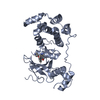
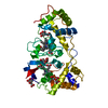
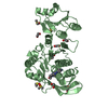
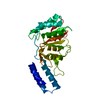
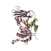
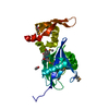
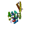
Crystal structure of sugar ABC transporter from Yersinia enterocolitica subsp. enterocolitica 8081
Components
Putative sugar ABC transporter
Keywords
SUGAR BINDING PROTEIN / ABC transporter / Methylthioribose-binding protein / Structural Genomics / Center for Structural Genomics of Infectious Diseases / CSGID
Function / homology
: / Periplasmic binding protein / Periplasmic binding protein domain / Periplasmic binding protein-like I / outer membrane-bounded periplasmic space / 5-Se-methyl-5-seleno-alpha-D-ribofuranose / 5-Se-methyl-5-seleno-beta-D-ribofuranose / Putative sugar ABC transporter / Sugar ABC transporter
Protocol: SINGLE WAVELENGTH / Monochromatic (M) / Laue (L): M / Scattering type: x-ray
Radiation wavelength
Wavelength: 0.9794 Å / Relative weight: 1
Reflection
Resolution: 1.28→40 Å / Num. obs: 78779 / % possible obs: 99.2 % / Redundancy: 5.7 % / Rmerge(I) obs: 0.12 / Net I/σ(I): 20
Reflection shell
Resolution: 1.28→1.3 Å / Rmerge(I) obs: 0.8 / % possible all: 92.6
-
Processing
Software
Name
Version
Classification
REFMAC
5.8.0158
refinement
HKL-3000
datareduction
HKL-3000
datascaling
HKL-3000
phasing
Refinement
Resolution: 1.28→40 Å / Cor.coef. Fo:Fc: 0.985 / Cor.coef. Fo:Fc free: 0.975 / SU B: 1.625 / SU ML: 0.03 / Cross valid method: THROUGHOUT / ESU R: 0.044 / ESU R Free: 0.047 / Details: HYDROGENS HAVE BEEN ADDED IN THE RIDING POSITIONS
Rfactor
Num. reflection
% reflection
Selection details
Rfree
0.153
3342
5 %
RANDOM
Rwork
0.11
-
-
-
obs
0.112
63186
93.8 %
-
Solvent computation
Ion probe radii: 0.8 Å / Shrinkage radii: 0.8 Å / VDW probe radii: 1.2 Å
 Movie
Movie Controller

 Yorodumi
 Open data
 Basic information
 Components
 Keywords
 Function and homology information
 Yersinia enterocolitica subsp. enterocolitica 8081 (bacteria)
 X-RAY DIFFRACTION /
  Authors
 Citation
 Structure visualization
 Downloads & links
 Other downloads
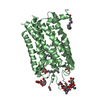


 PDBj
PDBj Assembly
 Type: D-saccharide, alpha linking / Mass: 227.117 Da / Num. of mol.: 1
 Type: D-saccharide, beta linking / Mass: 227.117 Da / Num. of mol.: 1
 Mass: 18.015 Da / Num. of mol.: 389 / Source method: isolated from a natural source / Formula: H2O
 Sample preparation
 / Beamline: 19-ID / Wavelength: 0.9794 Å
 Processing