 Movie
Movie Controller

[English] 日本語
 Yorodumi
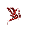
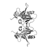
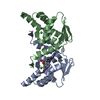
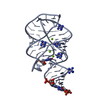
- PDB-4u2m: Crystal structure of a complex of the Miz1- and BCL6 POZ domains. -
+ Open data

- Basic information
| Entry | Database: PDB / ID: 4u2m | ||||||
|---|---|---|---|---|---|---|---|
| Title | Crystal structure of a complex of the Miz1- and BCL6 POZ domains. | ||||||
 Components Components | Zinc finger and BTB domain-containing protein 17,B-cell lymphoma 6 protein | ||||||
 Keywords Keywords | TRANSCRIPTION / POZ domain / BTB domain | ||||||
| Function / homology |  Function and homology information Function and homology informationXBP1(S) activates chaperone genes / regulation of memory T cell differentiation / negative regulation of mitotic cell cycle DNA replication / intronic transcription regulatory region sequence-specific DNA binding / isotype switching to IgE isotypes / negative regulation of plasma cell differentiation / negative regulation of T-helper 2 cell differentiation / negative regulation of isotype switching to IgE isotypes / negative regulation of mast cell cytokine production / regulation of germinal center formation ...XBP1(S) activates chaperone genes / regulation of memory T cell differentiation / negative regulation of mitotic cell cycle DNA replication / intronic transcription regulatory region sequence-specific DNA binding / isotype switching to IgE isotypes / negative regulation of plasma cell differentiation / negative regulation of T-helper 2 cell differentiation / negative regulation of isotype switching to IgE isotypes / negative regulation of mast cell cytokine production / regulation of germinal center formation / negative regulation of mononuclear cell proliferation / germinal center formation / plasma cell differentiation / paraspeckles / regulation of immune system process / type 2 immune response / negative regulation of Rho protein signal transduction / pyramidal neuron differentiation / T-helper 2 cell differentiation / positive regulation of regulatory T cell differentiation / negative regulation of B cell apoptotic process / positive regulation of cell motility / G1 to G0 transition / FOXO-mediated transcription of cell death genes / regulation of T cell proliferation / negative regulation of cellular senescence / negative regulation of cell-matrix adhesion / TP53 regulates transcription of several additional cell death genes whose specific roles in p53-dependent apoptosis remain uncertain / negative regulation of cell cycle / B cell proliferation / regulation of cell differentiation / negative regulation of Notch signaling pathway / regulation of immune response / core promoter sequence-specific DNA binding / erythrocyte development / Rho protein signal transduction / positive regulation of B cell proliferation / positive regulation of neuron differentiation / regulation of cytokine production / cell-matrix adhesion / transcription corepressor binding / cell motility / protein-DNA complex / chromatin DNA binding / negative regulation of cell growth / DNA-binding transcription repressor activity, RNA polymerase II-specific / cell morphogenesis / transcription coactivator binding / sequence-specific double-stranded DNA binding / intracellular protein localization / regulation of cell population proliferation / heterochromatin formation / regulation of inflammatory response / actin cytoskeleton organization / transcription by RNA polymerase II / Interleukin-4 and Interleukin-13 signaling / DNA-binding transcription activator activity, RNA polymerase II-specific / DNA-binding transcription factor binding / spermatogenesis / sequence-specific DNA binding / RNA polymerase II cis-regulatory region sequence-specific DNA binding / positive regulation of apoptotic process / DNA-binding transcription factor activity / inflammatory response / negative regulation of cell population proliferation / negative regulation of DNA-templated transcription / chromatin binding / DNA damage response / nucleolus / Golgi apparatus / negative regulation of transcription by RNA polymerase II / DNA-templated transcription / positive regulation of transcription by RNA polymerase II / protein-containing complex / zinc ion binding / nucleoplasm / identical protein binding / nucleus Similarity search - Function | ||||||
| Biological species |  Homo sapiens (human) Homo sapiens (human) | ||||||
| Method |  X-RAY DIFFRACTION / SYNCHROTRON / MOLECULAR REPLACEMENT / molecular replacement / Resolution: 2.23 Å X-RAY DIFFRACTION / SYNCHROTRON / MOLECULAR REPLACEMENT / molecular replacement / Resolution: 2.23 Å | ||||||
 Authors Authors | Stead, M.A. / Wright, S.C. | ||||||
| Funding support |  United Kingdom, 1items United Kingdom, 1items
| ||||||
 Citation Citation | Journal: Acta Crystallogr.,Sect.F / Year: 2014 Title: Structures of heterodimeric POZ domains of Miz1/BCL6 and Miz1/NAC1. Authors: Stead, M.A. / Wright, S.C. | ||||||
| History |
|
- Structure visualization
| Structure viewer | Molecule: MolmilJmol/JSmol |
|---|
- Downloads & links
-Download
| PDBx/mmCIF format | 4u2m.cif.gz | 190.7 KB | Display | PDBx/mmCIF format |
|---|---|---|---|---|
| PDB format | pdb4u2m.ent.gz | 152.6 KB | Display | PDB format |
| PDBx/mmJSON format | 4u2m.json.gz | Tree view | PDBx/mmJSON format | |
| Others |  Other downloads Other downloads |
-Validation report
| Arichive directory | https://data.pdbj.org/pub/pdb/validation_reports/u2/4u2mftp://data.pdbj.org/pub/pdb/validation_reports/u2/4u2m | HTTPS FTP |
|---|
-Related structure data


-Links
 PDBj
PDBj


- Assembly
| Deposited unit | 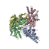
| ||||||||
|---|---|---|---|---|---|---|---|---|---|
| 1 | 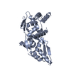
| ||||||||
| 2 | 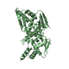
| ||||||||
| 3 | 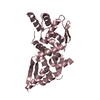
| ||||||||
| 4 | 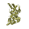
| ||||||||
| Unit cell |
| ||||||||
| Details | The biological unit is a dimer. There are four biological units in the asymmetric unit. |
-Components
| #1: Protein | Mass: 28327.648 Da / Num. of mol.: 4 Fragment: POZ domain, UNP residues 2-115,POZ domain, UNP residues 5-129 Source method: isolated from a genetically manipulated source Source: (gene. exp.) Homo sapiens (human)Gene: ZBTB17, MIZ1, ZNF151, ZNF60, BCL6, BCL5, LAZ3, ZBTB27, ZNF51 Plasmid: pGEX-6P-1 / Production host:  #2: Water | ChemComp-HOH / |  Mass: 18.015 Da / Num. of mol.: 163 / Source method: isolated from a natural source / Formula: H2O Mass: 18.015 Da / Num. of mol.: 163 / Source method: isolated from a natural source / Formula: H2O |
|---|
-Experimental details
-Experiment
| Experiment | Method: X-RAY DIFFRACTION / Number of used crystals: 1 |
|---|
- Sample preparation
| Crystal | Density Matthews: 2.38 Å3/Da / Density % sol: 48.24 % |
|---|---|
| Crystal grow | Temperature: 291 K / Method: vapor diffusion, hanging drop / pH: 4.5 Details: 4 % PEG 3350, 3 % 2-propanol, 0.2 M ammonium citrate pH 4.5 |
-Data collection
| Diffraction | Mean temperature: 100 K | |||||||||||||||||||||||||||
|---|---|---|---|---|---|---|---|---|---|---|---|---|---|---|---|---|---|---|---|---|---|---|---|---|---|---|---|---|
| Diffraction source | Source: SYNCHROTRON / Site: Diamond / Beamline: I04 / Wavelength: 0.97949 Å | |||||||||||||||||||||||||||
| Detector | Type: PSI PILATUS 6M / Detector: PIXEL / Date: Apr 25, 2014 | |||||||||||||||||||||||||||
| Radiation | Monochromator: DOUBLE CRYSTAL / Protocol: SINGLE WAVELENGTH / Monochromatic (M) / Laue (L): M / Scattering type: x-ray | |||||||||||||||||||||||||||
| Radiation wavelength | Wavelength: 0.97949 Å / Relative weight: 1 | |||||||||||||||||||||||||||
| Reflection | Resolution: 2.23→58.47 Å / Num. obs: 52962 / % possible obs: 99.3 % / Redundancy: 4.1 % / CC1/2: 0.997 / Rmerge(I) obs: 0.046 / Rpim(I) all: 0.025 / Net I/σ(I): 19 / Num. measured all: 215105 | |||||||||||||||||||||||||||
| Reflection shell | Diffraction-ID: 1 / Rejects: _
|
-Phasing
| Phasing | Method: molecular replacement | |||||||||
|---|---|---|---|---|---|---|---|---|---|---|
| Phasing MR | Model details: Phaser MODE: MR_AUTO
|
- Processing
| Software |
| |||||||||||||||||||||||||||||||||||||||||||||||||||||||||||||||||||||||||||
|---|---|---|---|---|---|---|---|---|---|---|---|---|---|---|---|---|---|---|---|---|---|---|---|---|---|---|---|---|---|---|---|---|---|---|---|---|---|---|---|---|---|---|---|---|---|---|---|---|---|---|---|---|---|---|---|---|---|---|---|---|---|---|---|---|---|---|---|---|---|---|---|---|---|---|---|---|
| Refinement | Method to determine structure: MOLECULAR REPLACEMENT / Resolution: 2.23→58.47 Å / Cor.coef. Fo:Fc: 0.948 / Cor.coef. Fo:Fc free: 0.932 / WRfactor Rfree: 0.227 / WRfactor Rwork: 0.198 / FOM work R set: 0.8008 / SU B: 6.899 / SU ML: 0.169 / SU R Cruickshank DPI: 0.2853 / SU Rfree: 0.209 / Cross valid method: THROUGHOUT / σ(F): 0 / ESU R: 0.285 / ESU R Free: 0.209 / Stereochemistry target values: MAXIMUM LIKELIHOOD Details: HYDROGENS HAVE BEEN ADDED IN THE RIDING POSITIONS U VALUES : REFINED INDIVIDUALLY
| |||||||||||||||||||||||||||||||||||||||||||||||||||||||||||||||||||||||||||
| Solvent computation | Ion probe radii: 0.8 Å / Shrinkage radii: 0.8 Å / VDW probe radii: 1.2 Å / Solvent model: MASK | |||||||||||||||||||||||||||||||||||||||||||||||||||||||||||||||||||||||||||
| Displacement parameters | Biso max: 112.85 Å2 / Biso mean: 46.076 Å2 / Biso min: 24.02 Å2
| |||||||||||||||||||||||||||||||||||||||||||||||||||||||||||||||||||||||||||
| Refinement step | Cycle: final / Resolution: 2.23→58.47 Å
| |||||||||||||||||||||||||||||||||||||||||||||||||||||||||||||||||||||||||||
| Refine LS restraints |
| |||||||||||||||||||||||||||||||||||||||||||||||||||||||||||||||||||||||||||
| LS refinement shell | Resolution: 2.23→2.288 Å / Total num. of bins used: 20
|
