endopeptidase La / ATP-dependent peptidase activity / protein quality control for misfolded or incompletely synthesized proteins / cellular response to heat / sequence-specific DNA binding / serine-type endopeptidase activity / ATP hydrolysis activity / ATP binding / metal ion binding / identical protein binding / cytoplasm Similarity search - Function
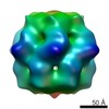
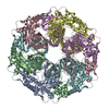
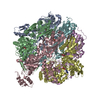
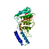
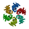
Journal: Structure / Year: 2016 Title: Structural Basis for the Magnesium-Dependent Activation and Hexamerization of the Lon AAA+ Protease. Authors: Shih-Chieh Su / Chien-Chu Lin / Hui-Chung Tai / Mu-Yueh Chang / Meng-Ru Ho / C Satheesan Babu / Jiahn-Haur Liao / Shih-Hsiung Wu / Yuan-Chih Chang / Carmay Lim / Chung-I Chang / Abstract: The Lon AAA+ protease (LonA) plays important roles in protein homeostasis and regulation of diverse biological processes. LonA behaves as a homomeric hexamer in the presence of magnesium (Mg(2+)) and ...The Lon AAA+ protease (LonA) plays important roles in protein homeostasis and regulation of diverse biological processes. LonA behaves as a homomeric hexamer in the presence of magnesium (Mg(2+)) and performs ATP-dependent proteolysis. However, it is also found that LonA can carry out Mg(2+)-dependent degradation of unfolded protein substrate in an ATP-independent manner. Here we show that in the presence of Mg(2+) LonA forms a non-secluded hexameric barrel with prominent openings, which explains why Mg(2+)-activated LonA can operate as a diffusion-based chambered protease to degrade unstructured protein and peptide substrates efficiently in the absence of ATP. A 1.85 Å crystal structure of Mg(2+)-activated protease domain reveals Mg(2+)-dependent remodeling of a substrate-binding loop and a potential metal-binding site near the Ser-Lys catalytic dyad, supported by biophysical binding assays and molecular dynamics simulations. Together, these findings reveal the specific roles of Mg(2+) in the molecular assembly and activation of LonA.
Resolution: 1.85→1.92 Å / Mean I/σ(I) obs: 4.629 / % possible all: 100
-
Processing
Software
Name
Version
Classification
REFMAC
5.8.0073
refinement
HKL-2000
dataprocessing
HKL-2000
datascaling
Coot
modelbuilding
Refinement
Resolution: 1.85→19.85 Å / Cor.coef. Fo:Fc: 0.956 / Cor.coef. Fo:Fc free: 0.948 / SU B: 2.137 / SU ML: 0.065 / Cross valid method: THROUGHOUT / ESU R: 0.101 / ESU R Free: 0.098 / Stereochemistry target values: MAXIMUM LIKELIHOOD / Details: HYDROGENS HAVE BEEN ADDED IN THE RIDING POSITIONS
Rfactor
Num. reflection
% reflection
Selection details
Rfree
0.20094
1924
4.9 %
RANDOM
Rwork
0.17714
-
-
-
obs
0.17835
37301
99.8 %
-
Solvent computation
Ion probe radii: 0.8 Å / Shrinkage radii: 0.8 Å / VDW probe radii: 1.2 Å / Solvent model: MASK
 Movie
Movie Controller

 Yorodumi
 Open data
 Basic information
 Components
 Keywords
 Function and homology information
 Meiothermus taiwanensis (bacteria)
 X-RAY DIFFRACTION /
  Authors
 Taiwan, 1items
  Citation
 Structure visualization
 Downloads & links
 Other downloads
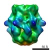


 PDBj
PDBj
 Assembly
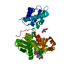
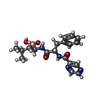


 Mass: 384.237 Da / Num. of mol.: 1 / Source method: obtained synthetically / Formula: C19H25BN4O4 / Comment: medication*YM
 Mass: 24.305 Da / Num. of mol.: 1 / Source method: obtained synthetically / Formula: Mg
 Mass: 106.120 Da / Num. of mol.: 1 / Source method: obtained synthetically / Formula: C4H10O3
 Mass: 192.124 Da / Num. of mol.: 1 / Source method: obtained synthetically / Formula: C6H8O7
 Sample preparation
 Processing