 Movie
Movie Controller


+ Open data

- Basic information
| Entry | Database: EMDB / ID: EMD-2697 | |||||||||
|---|---|---|---|---|---|---|---|---|---|---|
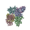
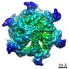
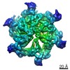
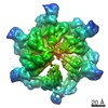
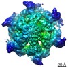
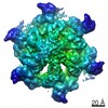
| Title | Cryo Electron Microscopy of GMPCPP-microtubules structure | |||||||||
 Map data Map data | 3D structure of GMPCPP-microtubules | |||||||||
 Sample Sample |
| |||||||||
 Keywords Keywords | Microtubule / tubuiln / GMPCPP / GTP-state structure / cryo-EM / microtubule stabilization / microtubule polymerization | |||||||||
| Function / homology |  Function and homology information Function and homology informationstructural constituent of cytoskeleton / microtubule cytoskeleton organization / neuron migration / mitotic cell cycle / microtubule / Hydrolases; Acting on acid anhydrides; Acting on GTP to facilitate cellular and subcellular movement / hydrolase activity / GTPase activity / GTP binding / metal ion binding / cytoplasm Similarity search - Function | |||||||||
| Biological species |  | |||||||||
| Method | single particle reconstruction / cryo EM / Resolution: 8.9 Å | |||||||||
 Authors Authors | Yajima H / Ogura T / Nitta R / Okada Y / Sato C / Hirokawa N | |||||||||
 Citation Citation | Journal: J Cell Biol / Year: 2012 Title: Conformational changes in tubulin in GMPCPP and GDP-taxol microtubules observed by cryoelectron microscopy. Authors: Hiroaki Yajima / Toshihiko Ogura / Ryo Nitta / Yasushi Okada / Chikara Sato / Nobutaka Hirokawa /  Abstract: Microtubules are dynamic polymers that stochastically switch between growing and shrinking phases. Microtubule dynamics are regulated by guanosine triphosphate (GTP) hydrolysis by β-tubulin, but the ...Microtubules are dynamic polymers that stochastically switch between growing and shrinking phases. Microtubule dynamics are regulated by guanosine triphosphate (GTP) hydrolysis by β-tubulin, but the mechanism of this regulation remains elusive because high-resolution microtubule structures have only been revealed for the guanosine diphosphate (GDP) state. In this paper, we solved the cryoelectron microscopy (cryo-EM) structure of microtubule stabilized with a GTP analogue, guanylyl 5'-α,β-methylenediphosphonate (GMPCPP), at 8.8-Å resolution by developing a novel cryo-EM image reconstruction algorithm. In contrast to the crystal structures of GTP-bound tubulin relatives such as γ-tubulin and bacterial tubulins, significant changes were detected between GMPCPP and GDP-taxol microtubules at the contacts between tubulins both along the protofilament and between neighboring protofilaments, contributing to the stability of the microtubule. These findings are consistent with the structural plasticity or lattice model and suggest the structural basis not only for the regulatory mechanism of microtubule dynamics but also for the recognition of the nucleotide state of the microtubule by several microtubule-binding proteins, such as EB1 or kinesin. | |||||||||
| History |
|
- Structure visualization
| Movie |
Movie viewer |
|---|---|
| Structure viewer | EM map: SurfViewMolmilJmol/JSmol |
| Supplemental images |

- Downloads & links
-EMDB archive
| Map data | emd_2697.map.gz | 125.8 KB | EMDB map data format | |
|---|---|---|---|---|
| Header (meta data) | emd-2697-v30.xmlemd-2697.xml | 12.5 KB 12.5 KB | Display Display | EMDB header |
| Images | EMD-2697_image_ver2.tif | 817.9 KB | ||
| Archive directory |  http://ftp.pdbj.org/pub/emdb/structures/EMD-2697ftp://ftp.pdbj.org/pub/emdb/structures/EMD-2697 http://ftp.pdbj.org/pub/emdb/structures/EMD-2697ftp://ftp.pdbj.org/pub/emdb/structures/EMD-2697 | HTTPS FTP |
-Related structure data
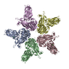
| Related structure data | 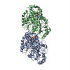 3j7iMC M: atomic model generated by this map C: citing same article ( |
|---|---|
| Similar structure data |

-Links
| EMDB pages | EMDB (EBI/PDBe) / EMDataResource |
|---|---|
| Related items in Molecule of the Month |


-Map
| File | Download / File: emd_2697.map.gz / Format: CCP4 / Size: 631.8 KB / Type: IMAGE STORED AS FLOATING POINT NUMBER (4 BYTES) | ||||||||||||||||||||||||||||||||||||||||||||||||||||||||||||||||||||
|---|---|---|---|---|---|---|---|---|---|---|---|---|---|---|---|---|---|---|---|---|---|---|---|---|---|---|---|---|---|---|---|---|---|---|---|---|---|---|---|---|---|---|---|---|---|---|---|---|---|---|---|---|---|---|---|---|---|---|---|---|---|---|---|---|---|---|---|---|---|
| Annotation | 3D structure of GMPCPP-microtubules | ||||||||||||||||||||||||||||||||||||||||||||||||||||||||||||||||||||
| Projections & slices | Image control
Images are generated by Spider. generated in cubic-lattice coordinate | ||||||||||||||||||||||||||||||||||||||||||||||||||||||||||||||||||||
| Voxel size | X=Y=Z: 2.5 Å | ||||||||||||||||||||||||||||||||||||||||||||||||||||||||||||||||||||
| Density |
| ||||||||||||||||||||||||||||||||||||||||||||||||||||||||||||||||||||
| Symmetry | Space group: 1 | ||||||||||||||||||||||||||||||||||||||||||||||||||||||||||||||||||||
| Details | EMDB XML:
CCP4 map header:
| ||||||||||||||||||||||||||||||||||||||||||||||||||||||||||||||||||||
 Z (Sec.)
 Y (Row.)
 X (Col.)


-Supplemental data
- Sample components
-Entire : GMPCPP-bound (GTP-state) microtubule
| Entire | Name: GMPCPP-bound (GTP-state) microtubule |
|---|---|
| Components |
|
-Supramolecule #1000: GMPCPP-bound (GTP-state) microtubule
| Supramolecule | Name: GMPCPP-bound (GTP-state) microtubule / type: sample / ID: 1000 / Oligomeric state: Alpha- and beta-tubulin heterodimer / Number unique components: 2 |
|---|---|
| Molecular weight | Experimental: 110 KDa / Theoretical: 110 KDa |
-Macromolecule #1: tubulin alpha chain
| Macromolecule | Name: tubulin alpha chain / type: protein_or_peptide / ID: 1 / Name.synonym: alpha tubulin Details: GTP binds on intra-tubulin dimer (between alpha- and beta-tubulin). Number of copies: 1 / Oligomeric state: hetero dimer / Recombinant expression: No |
|---|---|
| Source (natural) | Organism: |
| Molecular weight | Experimental: 55 KDa / Theoretical: 55 KDa |
| Sequence | UniProtKB: Tubulin alpha-1A chain |
-Macromolecule #2: tubulin beta chain
| Macromolecule | Name: tubulin beta chain / type: protein_or_peptide / ID: 2 / Name.synonym: beta tubulin Details: almost every beta-tubulin is bound to GMPCPP (>90% occupancy). without taxol. Number of copies: 1 / Oligomeric state: hetero dimer / Recombinant expression: No |
|---|---|
| Source (natural) | Organism: |
| Molecular weight | Experimental: 55 KDa / Theoretical: 55 KDa |
| Sequence | UniProtKB: Tubulin beta chain |
-Experimental details
-Structure determination
| Method | cryo EM |
|---|---|
 Processing Processing | single particle reconstruction |
| Aggregation state | particle |
-Sample preparation
| Concentration | 0.25 mg/mL |
|---|---|
| Buffer | pH: 6.8 Details: 100 mM Pipes, pH 6.8, adjusted by KOH, 1 mM EGTA, 1 mM MgCl2, 0.6 mM GMPCPP, 5% DMSO |
| Grid | Details: 300 mesh copper grid with thin holey carbon support, glow discharged in amylamine atmosphere |
| Vitrification | Cryogen name: ETHANE / Chamber temperature: 88 K / Instrument: LEICA KF80 Method: A 5-uL drop of the polymerized microtubules was placed onto a glow-discharged holey carbon film on a copper mesh grid. After 30 seconds, this solution was absorbed by filter paper and quickly ...Method: A 5-uL drop of the polymerized microtubules was placed onto a glow-discharged holey carbon film on a copper mesh grid. After 30 seconds, this solution was absorbed by filter paper and quickly replaced with an 8-uL drop of the same buffer without microtubules. Immediately after blotting this drop with filter papers from both grid sides for 5 seconds, the grid was plunge frozen in liquid ethane. |
- Electron microscopy
| Microscope | JEOL 2010F |
|---|---|
| Temperature | Min: 90 K / Max: 95 K / Average: 93 K |
| Alignment procedure | Legacy - Astigmatism: Objective lens astigmatism was corrected at 100,000 times magnification |
| Date | Jun 12, 2010 |
| Image recording | Category: FILM / Film or detector model: KODAK SO-163 FILM / Digitization - Scanner: OTHER / Digitization - Sampling interval: 10 µm / Number real images: 350 / Average electron dose: 10.00 e/Å2 / Bits/pixel: 16 |
| Tilt angle min | 0 |
| Tilt angle max | 0 |
| Electron beam | Acceleration voltage: 200 kV / Electron source:  FIELD EMISSION GUN FIELD EMISSION GUN |
| Electron optics | Calibrated magnification: 40000 / Illumination mode: FLOOD BEAM / Imaging mode: BRIGHT FIELD / Cs: 3.3 mm / Nominal defocus max: 2.8 µm / Nominal defocus min: 1.3 µm / Nominal magnification: 40000 |
| Sample stage | Specimen holder: Gatan 626 Single Tilt Liquid Nitrogen Cryo Transfer Holder Specimen holder model: GATAN LIQUID NITROGEN |
-Image processing
| Details | The particles were selected along individual microtubules using an automatic selection program. |
|---|---|
| CTF correction | Details: Each filament |
| Final reconstruction | Applied symmetry - Point group: C1 (asymmetric) / Algorithm: OTHER / Resolution.type: BY AUTHOR / Resolution: 8.9 Å / Resolution method: OTHER / Software - Name: MATLAB, ImageV / Number images used: 320000 |
| Final two d classification | Number classes: 18 |
-Atomic model buiding 1
| Initial model | PDB ID: Chain - #0 - Chain ID: A / Chain - #1 - Chain ID: B |
|---|---|
| Software | Name: Chimera |
| Details | Initial local fitting was done before the domains were separately fitted by using Chimera. |
| Refinement | Space: REAL / Protocol: RIGID BODY FIT / Target criteria: Cross-correlation, Average map value |
| Output model | PDB-3j7i: |

