 Movie
Movie Controller


+ Open data

- Basic information
| Entry | Database: PDB / ID: 3j7i | ||||||
|---|---|---|---|---|---|---|---|
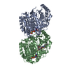
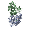
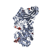
| Title | Structure of alpha- and beta- tubulin in GMPCPP-microtubules | ||||||
 Components Components |
| ||||||
 Keywords Keywords | STRUCTURAL PROTEIN / Microtubule / Tubulin / GTP-state structure / GMPCPP / Microtubule stabilaization / Micotubule polymerization | ||||||
| Function / homology |  Function and homology information Function and homology informationstructural constituent of cytoskeleton / microtubule cytoskeleton organization / neuron migration / mitotic cell cycle / microtubule / Hydrolases; Acting on acid anhydrides; Acting on GTP to facilitate cellular and subcellular movement / hydrolase activity / GTPase activity / GTP binding / metal ion binding / cytoplasm Similarity search - Function | ||||||
| Biological species |  | ||||||
| Method | ELECTRON MICROSCOPY / single particle reconstruction / cryo EM / Resolution: 8.9 Å | ||||||
 Authors Authors | Yajima, H. / Ogura, T. / Nitta, R. / Okada, Y. / Sato, C. / Hirokawa, N. | ||||||
 Citation Citation | Journal: J Cell Biol / Year: 2012 Title: Conformational changes in tubulin in GMPCPP and GDP-taxol microtubules observed by cryoelectron microscopy. Authors: Hiroaki Yajima / Toshihiko Ogura / Ryo Nitta / Yasushi Okada / Chikara Sato / Nobutaka Hirokawa /  Abstract: Microtubules are dynamic polymers that stochastically switch between growing and shrinking phases. Microtubule dynamics are regulated by guanosine triphosphate (GTP) hydrolysis by β-tubulin, but the ...Microtubules are dynamic polymers that stochastically switch between growing and shrinking phases. Microtubule dynamics are regulated by guanosine triphosphate (GTP) hydrolysis by β-tubulin, but the mechanism of this regulation remains elusive because high-resolution microtubule structures have only been revealed for the guanosine diphosphate (GDP) state. In this paper, we solved the cryoelectron microscopy (cryo-EM) structure of microtubule stabilized with a GTP analogue, guanylyl 5'-α,β-methylenediphosphonate (GMPCPP), at 8.8-Å resolution by developing a novel cryo-EM image reconstruction algorithm. In contrast to the crystal structures of GTP-bound tubulin relatives such as γ-tubulin and bacterial tubulins, significant changes were detected between GMPCPP and GDP-taxol microtubules at the contacts between tubulins both along the protofilament and between neighboring protofilaments, contributing to the stability of the microtubule. These findings are consistent with the structural plasticity or lattice model and suggest the structural basis not only for the regulatory mechanism of microtubule dynamics but also for the recognition of the nucleotide state of the microtubule by several microtubule-binding proteins, such as EB1 or kinesin. #1: Journal: To Be PublishedTitle: Mutual Conformational Changes of Kinesin and GTP-Microtubule Upon their Binding Authors: Yajima, H. / Ogura, T. / Nitta, R. / Okada, Y. / Sato, C. / Hirokawa, N. | ||||||
| History |
|
- Structure visualization
| Movie |
Movie viewer |
|---|---|
| Structure viewer | Molecule: MolmilJmol/JSmol |
- Downloads & links
-Download
| PDBx/mmCIF format | 3j7i.cif.gz | 180 KB | Display | PDBx/mmCIF format |
|---|---|---|---|---|
| PDB format | pdb3j7i.ent.gz | 137.7 KB | Display | PDB format |
| PDBx/mmJSON format | 3j7i.json.gz | Tree view | PDBx/mmJSON format | |
| Others |  Other downloads Other downloads |
-Validation report
| Arichive directory | https://data.pdbj.org/pub/pdb/validation_reports/j7/3j7iftp://data.pdbj.org/pub/pdb/validation_reports/j7/3j7i | HTTPS FTP |
|---|
-Related structure data
| Related structure data |  2697MC M: map data used to model this data C: citing same article ( |
|---|---|
| Similar structure data |


-Links
 PDBj
PDBj


- Assembly
| Deposited unit | 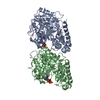
|
|---|---|
| 1 |
|
-Components
| #1: Protein | Mass: 50107.238 Da / Num. of mol.: 1 / Source method: isolated from a natural source / Source: (natural) | ||||
|---|---|---|---|---|---|
| #2: Protein | Mass: 49907.770 Da / Num. of mol.: 1 / Source method: isolated from a natural source / Source: (natural) | ||||
| #3: Chemical |   Mass: 24.305 Da / Num. of mol.: 2 / Source method: obtained synthetically / Formula: Mg Mass: 24.305 Da / Num. of mol.: 2 / Source method: obtained synthetically / Formula: Mg#4: Chemical |   Mass: 523.180 Da / Num. of mol.: 2 / Source method: obtained synthetically / Formula: C10H16N5O14P3 / Comment: GTP, energy-carrying molecule*YM Mass: 523.180 Da / Num. of mol.: 2 / Source method: obtained synthetically / Formula: C10H16N5O14P3 / Comment: GTP, energy-carrying molecule*YMSequence details | THIS SEQUENCE IS NATURAL VARIANT. | |
-Experimental details
-Experiment
| Experiment | Method: ELECTRON MICROSCOPY |
|---|---|
| EM experiment | Aggregation state: HELICAL ARRAY / 3D reconstruction method: single particle reconstruction |
- Sample preparation
| Component | Name: GMPCPP-microtubules / Type: COMPLEX |
|---|---|
| Buffer solution | pH: 6.8 |
| Specimen | Embedding applied: NO / Shadowing applied: NO / Staining applied: NO / Vitrification applied: YES |
| Vitrification | Instrument: LEICA KF80 / Cryogen name: ETHANE |
- Electron microscopy imaging
| Microscopy | Model: JEOL 2010F / Date: Jan 1, 2010 |
|---|---|
| Electron gun | Electron source:  FIELD EMISSION GUN / Accelerating voltage: 200 kV / Illumination mode: FLOOD BEAM FIELD EMISSION GUN / Accelerating voltage: 200 kV / Illumination mode: FLOOD BEAM |
| Electron lens | Mode: BRIGHT FIELD / Nominal magnification: 40000 X / Calibrated magnification: 40000 X / Nominal defocus max: 2800 nm / Nominal defocus min: 1300 nm / Cs: 3.3 mm |
| Specimen holder | Temperature: 88 K / Tilt angle max: 0 ° / Tilt angle min: 0 ° |
| Image recording | Electron dose: 10 e/Å2 / Film or detector model: KODAK SO-163 FILM |
- Processing
| EM software |
| ||||||||||||||||
|---|---|---|---|---|---|---|---|---|---|---|---|---|---|---|---|---|---|
| CTF correction | Details: Each Filament | ||||||||||||||||
| Symmetry | Point symmetry: C1 (asymmetric) | ||||||||||||||||
| 3D reconstruction | Method: Single Particle / Resolution: 8.9 Å / Num. of particles: 320000 / Nominal pixel size: 2.5 Å / Actual pixel size: 2.5 Å / Num. of class averages: 18 / Symmetry type: POINT | ||||||||||||||||
| Atomic model building | Protocol: RIGID BODY FIT / Space: REAL / Target criteria: Cross-correlation, Average map value Details: METHOD--Local refinement, Domain fitting REFINEMENT PROTOCOL--Rigid body refinement DETAILS--Initial local fitting was done before the domains were separately fitted by using Chimera. | ||||||||||||||||
| Atomic model building | PDB-ID: 1JFF Accession code: 1JFF / Source name: PDB / Type: experimental model | ||||||||||||||||
| Refinement step | Cycle: LAST
|

