 Movie
Movie Controller

[English] 日本語
 Yorodumi
- PDB-4ypl: Crystal structure of a hexameric LonA protease bound to three ADPs -
+ Open data

- Basic information
| Entry | Database: PDB / ID: 4ypl | ||||||
|---|---|---|---|---|---|---|---|
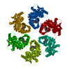
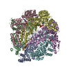
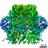
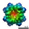
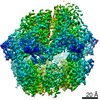
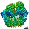
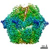
| Title | Crystal structure of a hexameric LonA protease bound to three ADPs | ||||||
 Components Components | Lon protease | ||||||
 Keywords Keywords | HYDROLASE / Lon protease / ADP / MMH8709 / Inhibitor / AAA+ domain | ||||||
| Function / homology |  Function and homology information Function and homology informationendopeptidase La / ATP-dependent peptidase activity / protein quality control for misfolded or incompletely synthesized proteins / cellular response to heat / sequence-specific DNA binding / serine-type endopeptidase activity / ATP hydrolysis activity / ATP binding / metal ion binding / identical protein binding / cytoplasm Similarity search - Function | ||||||
| Biological species |  Meiothermus taiwanensis (bacteria) Meiothermus taiwanensis (bacteria) | ||||||
| Method |  X-RAY DIFFRACTION / SYNCHROTRON / MOLECULAR REPLACEMENT / Resolution: 3.45 Å X-RAY DIFFRACTION / SYNCHROTRON / MOLECULAR REPLACEMENT / Resolution: 3.45 Å | ||||||
 Authors Authors | Lin, C.-C. / Chang, C.-I. | ||||||
 Citation Citation | Journal: Structure / Year: 2016 Title: Structural Insights into the Allosteric Operation of the Lon AAA+ Protease Authors: Lin, C.-C. / Su, S.-C. / Su, M.-Y. / Liang, P.-H. / Feng, C.-C. / Wu, S.-H. / Chang, C.-I. | ||||||
| History |
|
- Structure visualization
| Structure viewer | Molecule: MolmilJmol/JSmol |
|---|
- Downloads & links
-Download
| PDBx/mmCIF format | 4ypl.cif.gz | 616.7 KB | Display | PDBx/mmCIF format |
|---|---|---|---|---|
| PDB format | pdb4ypl.ent.gz | 511.1 KB | Display | PDB format |
| PDBx/mmJSON format | 4ypl.json.gz | Tree view | PDBx/mmJSON format | |
| Others |  Other downloads Other downloads |
-Validation report
| Arichive directory | https://data.pdbj.org/pub/pdb/validation_reports/yp/4yplftp://data.pdbj.org/pub/pdb/validation_reports/yp/4ypl | HTTPS FTP |
|---|
-Related structure data
| Related structure data | 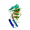 4ypnSC 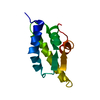 1qzmS 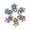 1rr9S S: Starting model for refinement C: citing same article ( |
|---|---|
| Similar structure data |


-Links
 PDBj
PDBj
- Assembly
| Deposited unit | 
| ||||||||
|---|---|---|---|---|---|---|---|---|---|
| 1 |
| ||||||||
| Unit cell |
|
-Components
| #1: Protein | Mass: 61329.273 Da / Num. of mol.: 6 Fragment: AAA+ domain, protease domain, UNP residues 242-793 Mutation: E423Q Source method: isolated from a genetically manipulated source Source: (gene. exp.) Meiothermus taiwanensis (bacteria) / Gene: lonA1, lon / Production host: #2: Chemical | ChemComp-4KZ /   Mass: 418.253 Da / Num. of mol.: 6 / Source method: obtained synthetically / Formula: C22H23BN4O4 Mass: 418.253 Da / Num. of mol.: 6 / Source method: obtained synthetically / Formula: C22H23BN4O4#3: Chemical |   Mass: 427.201 Da / Num. of mol.: 3 / Source method: obtained synthetically / Formula: C10H15N5O10P2 / Comment: ADP, energy-carrying molecule*YM Mass: 427.201 Da / Num. of mol.: 3 / Source method: obtained synthetically / Formula: C10H15N5O10P2 / Comment: ADP, energy-carrying molecule*YMHas protein modification | Y | |
|---|
-Experimental details
-Experiment
| Experiment | Method: X-RAY DIFFRACTION / Number of used crystals: 1 |
|---|
- Sample preparation
| Crystal | Density Matthews: 2.63 Å3/Da / Density % sol: 53.22 % |
|---|---|
| Crystal grow | Temperature: 295 K / Method: evaporation / pH: 8.7 Details: 0.1 M Tris-HCl pH 8.7, 0.1 M CaCl2, 10-13% PEG 3350 |
-Data collection
| Diffraction | Mean temperature: 200 K |
|---|---|
| Diffraction source | Source: SYNCHROTRON / Site: SPring-8  / Beamline: BL44XU / Wavelength: 0.9 Å / Beamline: BL44XU / Wavelength: 0.9 Å |
| Detector | Type: RAYONIX MX300HE / Detector: CCD / Date: Nov 22, 2014 |
| Radiation | Protocol: SINGLE WAVELENGTH / Monochromatic (M) / Laue (L): M / Scattering type: x-ray |
| Radiation wavelength | Wavelength: 0.9 Å / Relative weight: 1 |
| Reflection | Resolution: 3.45→50 Å / Num. all: 52050 / Num. obs: 45968 / % possible obs: 99.5 % / Observed criterion σ(I): 2 / Redundancy: 5.6 % / Net I/av σ(I): 13.22 / Net I/σ(I): 0.168 |
| Reflection shell | Resolution: 3.45→3.47 Å / Redundancy: 5.4 % / Mean I/σ(I) obs: 2.53 / % possible all: 99.6 |
- Processing
| Software |
| ||||||||||||||||||||||||||||||||||||||||||||||||||||||||||||||||||||||||||||||||||||||||||||||||||||||||||||||||||||||||||||||||||||||||||||||||||||||||||||||||||||||||||||||||||||||
|---|---|---|---|---|---|---|---|---|---|---|---|---|---|---|---|---|---|---|---|---|---|---|---|---|---|---|---|---|---|---|---|---|---|---|---|---|---|---|---|---|---|---|---|---|---|---|---|---|---|---|---|---|---|---|---|---|---|---|---|---|---|---|---|---|---|---|---|---|---|---|---|---|---|---|---|---|---|---|---|---|---|---|---|---|---|---|---|---|---|---|---|---|---|---|---|---|---|---|---|---|---|---|---|---|---|---|---|---|---|---|---|---|---|---|---|---|---|---|---|---|---|---|---|---|---|---|---|---|---|---|---|---|---|---|---|---|---|---|---|---|---|---|---|---|---|---|---|---|---|---|---|---|---|---|---|---|---|---|---|---|---|---|---|---|---|---|---|---|---|---|---|---|---|---|---|---|---|---|---|---|---|---|---|
| Refinement | Method to determine structure: MOLECULAR REPLACEMENT Starting model: 4YPN, 1QZM and 1RR9 Resolution: 3.45→30 Å / Cor.coef. Fo:Fc: 0.875 / Cor.coef. Fo:Fc free: 0.841 / Cross valid method: THROUGHOUT / ESU R Free: 0.706 / Stereochemistry target values: MAXIMUM LIKELIHOOD / Details: HYDROGENS HAVE BEEN ADDED IN THE RIDING POSITIONS
| ||||||||||||||||||||||||||||||||||||||||||||||||||||||||||||||||||||||||||||||||||||||||||||||||||||||||||||||||||||||||||||||||||||||||||||||||||||||||||||||||||||||||||||||||||||||
| Solvent computation | Ion probe radii: 0.8 Å / Shrinkage radii: 0.8 Å / VDW probe radii: 1.2 Å / Solvent model: MASK | ||||||||||||||||||||||||||||||||||||||||||||||||||||||||||||||||||||||||||||||||||||||||||||||||||||||||||||||||||||||||||||||||||||||||||||||||||||||||||||||||||||||||||||||||||||||
| Displacement parameters | Biso mean: 66.574 Å2
| ||||||||||||||||||||||||||||||||||||||||||||||||||||||||||||||||||||||||||||||||||||||||||||||||||||||||||||||||||||||||||||||||||||||||||||||||||||||||||||||||||||||||||||||||||||||
| Refinement step | Cycle: LAST / Resolution: 3.45→30 Å
| ||||||||||||||||||||||||||||||||||||||||||||||||||||||||||||||||||||||||||||||||||||||||||||||||||||||||||||||||||||||||||||||||||||||||||||||||||||||||||||||||||||||||||||||||||||||
| Refine LS restraints |
|