[English] 日本語
 Yorodumi
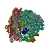
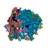
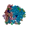
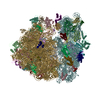
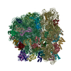
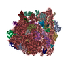
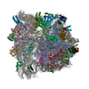
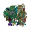
- PDB-4v6k: Structural insights into cognate vs. near-cognate discrimination ... -
+ Open data
- Basic information
| Entry | Database: PDB / ID: 4v6k | |||||||||||||||
|---|---|---|---|---|---|---|---|---|---|---|---|---|---|---|---|---|
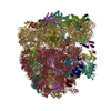
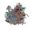
| Title | Structural insights into cognate vs. near-cognate discrimination during decoding. | |||||||||||||||
 Components Components |
| |||||||||||||||
 Keywords Keywords | RIBOSOME / translation / ternary complex / tRNA incorporation / cryoEM / near-cognate | |||||||||||||||
| Function / homology |  Function and homology information Function and homology informationguanyl-nucleotide exchange factor complex / guanosine tetraphosphate binding / negative regulation of cytoplasmic translational initiation / stringent response / ornithine decarboxylase inhibitor activity / transcription antitermination factor activity, RNA binding / misfolded RNA binding / Group I intron splicing / RNA folding / translational elongation ...guanyl-nucleotide exchange factor complex / guanosine tetraphosphate binding / negative regulation of cytoplasmic translational initiation / stringent response / ornithine decarboxylase inhibitor activity / transcription antitermination factor activity, RNA binding / misfolded RNA binding / Group I intron splicing / RNA folding / translational elongation / transcriptional attenuation / translation elongation factor activity / positive regulation of ribosome biogenesis / endoribonuclease inhibitor activity / RNA-binding transcription regulator activity / translational termination / negative regulation of cytoplasmic translation / four-way junction DNA binding / DnaA-L2 complex / translation repressor activity / negative regulation of translational initiation / regulation of mRNA stability / negative regulation of DNA-templated DNA replication initiation / mRNA regulatory element binding translation repressor activity / assembly of large subunit precursor of preribosome / positive regulation of RNA splicing / ribosome assembly / transcription elongation factor complex / cytosolic ribosome assembly / regulation of DNA-templated transcription elongation / DNA endonuclease activity / response to reactive oxygen species / transcription antitermination / translational initiation / regulation of cell growth / DNA-templated transcription termination / response to radiation / maintenance of translational fidelity / mRNA 5'-UTR binding / regulation of translation / ribosome biogenesis / large ribosomal subunit / ribosome binding / transferase activity / ribosomal small subunit biogenesis / ribosomal small subunit assembly / small ribosomal subunit / small ribosomal subunit rRNA binding / 5S rRNA binding / ribosomal large subunit assembly / cytosolic small ribosomal subunit / large ribosomal subunit rRNA binding / cytosolic large ribosomal subunit / cytoplasmic translation / tRNA binding / negative regulation of translation / rRNA binding / ribosome / structural constituent of ribosome / translation / response to antibiotic / GTPase activity / negative regulation of DNA-templated transcription / mRNA binding / GTP binding / DNA binding / RNA binding / zinc ion binding / membrane / plasma membrane / cytosol / cytoplasm Similarity search - Function | |||||||||||||||
| Biological species |     | |||||||||||||||
| Method | ELECTRON MICROSCOPY / single particle reconstruction / cryo EM / Resolution: 8.25 Å | |||||||||||||||
 Authors Authors | Agirrezabala, X. / Schreiner, E. / Trabuco, L.G. / Lei, J. / Ortiz-Meoz, R.F. / Schulten, K. / Green, R. / Frank, J. | |||||||||||||||
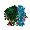
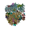
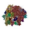
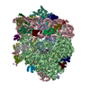
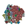
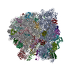
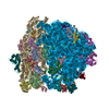
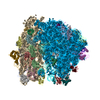
 Citation Citation |  Journal: EMBO J / Year: 2011 Journal: EMBO J / Year: 2011Title: Structural insights into cognate versus near-cognate discrimination during decoding. Authors: Xabier Agirrezabala / Eduard Schreiner / Leonardo G Trabuco / Jianlin Lei / Rodrigo F Ortiz-Meoz / Klaus Schulten / Rachel Green / Joachim Frank /  Abstract: The structural basis of the tRNA selection process is investigated by cryo-electron microscopy of ribosomes programmed with UGA codons and incubated with ternary complex (TC) containing the near- ...The structural basis of the tRNA selection process is investigated by cryo-electron microscopy of ribosomes programmed with UGA codons and incubated with ternary complex (TC) containing the near-cognate Trp-tRNA(Trp) in the presence of kirromycin. Going through more than 350 000 images and employing image classification procedures, we find ∼8% in which the TC is bound to the ribosome. The reconstructed 3D map provides a means to characterize the arrangement of the near-cognate aa-tRNA with respect to elongation factor Tu (EF-Tu) and the ribosome, as well as the domain movements of the ribosome. One of the interesting findings is that near-cognate tRNA's acceptor stem region is flexible and CCA end becomes disordered. The data bring direct structural insights into the induced-fit mechanism of decoding by the ribosome, as the analysis of the interactions between small and large ribosomal subunit, aa-tRNA and EF-Tu and comparison with the cognate case (UGG codon) offers clues on how the conformational signals conveyed to the GTPase differ in the two cases. | |||||||||||||||
| History |
|
- Structure visualization
| Movie |
 Movie viewer Movie viewer |
|---|---|
| Structure viewer | Molecule:  Molmil Molmil Jmol/JSmol Jmol/JSmol |
- Downloads & links
- Download
| PDBx/mmCIF format |  4v6k.cif.gz 4v6k.cif.gz | 3.4 MB | Display |  PDBx/mmCIF format PDBx/mmCIF format |
|---|---|---|---|---|
| PDB format |  pdb4v6k.ent.gz pdb4v6k.ent.gz | Display |  PDB format PDB format | |
| PDBx/mmJSON format |  4v6k.json.gz 4v6k.json.gz | Tree view |  PDBx/mmJSON format PDBx/mmJSON format | |
| Others |  Other downloads Other downloads |
-Validation report
| Summary document |  4v6k_validation.pdf.gz 4v6k_validation.pdf.gz | 1.3 MB | Display |  wwPDB validaton report wwPDB validaton report |
|---|---|---|---|---|
| Full document |  4v6k_full_validation.pdf.gz 4v6k_full_validation.pdf.gz | 1.8 MB | Display | |
| Data in XML |  4v6k_validation.xml.gz 4v6k_validation.xml.gz | 251.7 KB | Display | |
| Data in CIF |  4v6k_validation.cif.gz 4v6k_validation.cif.gz | 436.8 KB | Display | |
| Arichive directory |  https://data.pdbj.org/pub/pdb/validation_reports/v6/4v6k https://data.pdbj.org/pub/pdb/validation_reports/v6/4v6k ftp://data.pdbj.org/pub/pdb/validation_reports/v6/4v6k ftp://data.pdbj.org/pub/pdb/validation_reports/v6/4v6k | HTTPS FTP |
-Related structure data
| Related structure data |  1849MC  1850C  4v6lC M: map data used to model this data C: citing same article ( |
|---|---|
| Similar structure data |
- Links
Links
- Assembly
| Deposited unit | 
|
|---|---|
| 1 |
|
- Components
-Ribosomal RNA ... , 2 types, 2 molecules AAAB
| #1: RNA chain | Mass: 38790.090 Da / Num. of mol.: 1 / Source method: isolated from a natural source / Source: (natural)  |
|---|---|
| #2: RNA chain | Mass: 941813.562 Da / Num. of mol.: 1 / Source method: isolated from a natural source / Source: (natural)  |
+50S ribosomal protein ... , 31 types, 31 molecules ACADAEAFAGAHAIAJAKALAMANAOAPAQARASATAUAVAWAXAYAZAaAbAcAdAeAfAg
-RNA chain , 3 types, 4 molecules BABBBEBD
| #34: RNA chain | Mass: 499874.406 Da / Num. of mol.: 1 / Source method: isolated from a natural source / Source: (natural)  | ||
|---|---|---|---|
| #35: RNA chain | Mass: 24751.018 Da / Num. of mol.: 2 / Source method: isolated from a natural source / Source: (natural)  #37: RNA chain | | Mass: 7487.294 Da / Num. of mol.: 1 / Source method: isolated from a natural source / Source: (natural)  |
-Protein , 1 types, 1 molecules BC
| #36: Protein | Mass: 43239.297 Da / Num. of mol.: 1 / Source method: isolated from a natural source / Source: (natural)  |
|---|
-30S ribosomal protein ... , 20 types, 20 molecules BFBGBHBIBJBKBLBMBNBOBPBQBRBSBTBUBVBWBXBY
| #38: Protein | Mass: 26781.670 Da / Num. of mol.: 1 / Source method: isolated from a natural source / Source: (natural)  |
|---|---|
| #39: Protein | Mass: 26031.316 Da / Num. of mol.: 1 / Source method: isolated from a natural source / Source: (natural)  |
| #40: Protein | Mass: 23514.199 Da / Num. of mol.: 1 / Source method: isolated from a natural source / Source: (natural)  |
| #41: Protein | Mass: 17629.398 Da / Num. of mol.: 1 / Source method: isolated from a natural source / Source: (natural)  |
| #42: Protein | Mass: 15727.512 Da / Num. of mol.: 1 / Source method: isolated from a natural source / Source: (natural)  |
| #43: Protein | Mass: 20055.156 Da / Num. of mol.: 1 / Source method: isolated from a natural source / Source: (natural)  |
| #44: Protein | Mass: 14146.557 Da / Num. of mol.: 1 / Source method: isolated from a natural source / Source: (natural)  |
| #45: Protein | Mass: 14886.270 Da / Num. of mol.: 1 / Source method: isolated from a natural source / Source: (natural)  |
| #46: Protein | Mass: 11755.597 Da / Num. of mol.: 1 / Source method: isolated from a natural source / Source: (natural)  |
| #47: Protein | Mass: 13870.975 Da / Num. of mol.: 1 / Source method: isolated from a natural source / Source: (natural)  |
| #48: Protein | Mass: 13768.157 Da / Num. of mol.: 1 / Source method: isolated from a natural source / Source: (natural)  |
| #49: Protein | Mass: 13128.467 Da / Num. of mol.: 1 / Source method: isolated from a natural source / Source: (natural)  |
| #50: Protein | Mass: 11606.560 Da / Num. of mol.: 1 / Source method: isolated from a natural source / Source: (natural)  |
| #51: Protein | Mass: 10319.882 Da / Num. of mol.: 1 / Source method: isolated from a natural source / Source: (natural)  |
| #52: Protein | Mass: 9207.572 Da / Num. of mol.: 1 / Source method: isolated from a natural source / Source: (natural)  |
| #53: Protein | Mass: 9724.491 Da / Num. of mol.: 1 / Source method: isolated from a natural source / Source: (natural)  |
| #54: Protein | Mass: 9005.472 Da / Num. of mol.: 1 / Source method: isolated from a natural source / Source: (natural)  |
| #55: Protein | Mass: 10455.355 Da / Num. of mol.: 1 / Source method: isolated from a natural source / Source: (natural)  |
| #56: Protein | Mass: 9708.464 Da / Num. of mol.: 1 / Source method: isolated from a natural source / Source: (natural)  |
| #57: Protein | Mass: 8524.039 Da / Num. of mol.: 1 / Source method: isolated from a natural source / Source: (natural)  |
-Details
| Has protein modification | N |
|---|
-Experimental details
-Experiment
| Experiment | Method: ELECTRON MICROSCOPY |
|---|---|
| EM experiment | Aggregation state: PARTICLE / 3D reconstruction method: single particle reconstruction |
- Sample preparation
| Component |
| ||||||||||||
|---|---|---|---|---|---|---|---|---|---|---|---|---|---|
| Buffer solution | pH: 7.5 | ||||||||||||
| Specimen | Embedding applied: NO / Shadowing applied: NO / Staining applied: NO / Vitrification applied: YES | ||||||||||||
| Vitrification | Instrument: FEI VITROBOT MARK I / Cryogen name: NITROGEN |
- Electron microscopy imaging
| Experimental equipment |  Model: Tecnai F30 / Image courtesy: FEI Company |
|---|---|
| Microscopy | Model: FEI TECNAI F30 |
| Electron gun | Electron source:  FIELD EMISSION GUN / Accelerating voltage: 300 kV / Illumination mode: FLOOD BEAM FIELD EMISSION GUN / Accelerating voltage: 300 kV / Illumination mode: FLOOD BEAM |
| Electron lens | Mode: BRIGHT FIELD / Nominal magnification: 59000 X / Nominal defocus max: 4000 nm / Nominal defocus min: 1200 nm |
| Image recording | Electron dose: 20 e/Å2 / Film or detector model: TVIPS TEMCAM-F415 (4k x 4k) |
| Radiation | Protocol: SINGLE WAVELENGTH / Monochromatic (M) / Laue (L): M / Scattering type: x-ray |
| Radiation wavelength | Relative weight: 1 |
- Processing
| EM software |
| ||||||||||||
|---|---|---|---|---|---|---|---|---|---|---|---|---|---|
| Symmetry | Point symmetry: C1 (asymmetric) | ||||||||||||
| 3D reconstruction | Resolution: 8.25 Å / Num. of particles: 359223 / Symmetry type: POINT | ||||||||||||
| Atomic model building | Protocol: FLEXIBLE FIT / Space: REAL / Target criteria: RMSD < 0.1 A/ns / Details: METHOD--flexible fitting, MDFF | ||||||||||||
| Atomic model building | PDB-ID: 2I2V 2i2v Accession code: 2I2V / Source name: PDB / Type: experimental model | ||||||||||||
| Refinement step | Cycle: LAST
|
 Movie
Movie Controller



 PDBj
PDBj


