 Movie
Movie Controller

[English] 日本語
 Yorodumi
- PDB-7nhl: VgaA-LC, an antibiotic resistance ABCF, in complex with 70S ribos... -
+ Open data

- Basic information
| Entry | Database: PDB / ID: 7nhl | |||||||||||||||||||||
|---|---|---|---|---|---|---|---|---|---|---|---|---|---|---|---|---|---|---|---|---|---|---|
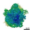
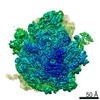
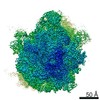
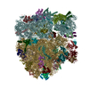
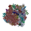
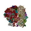
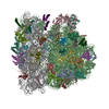
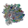
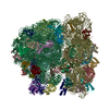
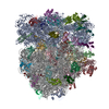
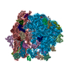
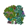
| Title | VgaA-LC, an antibiotic resistance ABCF, in complex with 70S ribosome from Staphylococcus aureus | |||||||||||||||||||||
 Components Components |
| |||||||||||||||||||||
 Keywords Keywords | RIBOSOME / Antibiotic resistance element / ABCF / antibiotic resistance / ATPase / protein synthesis | |||||||||||||||||||||
| Function / homology |  Function and homology information Function and homology informationlarge ribosomal subunit / transferase activity / ribosome biogenesis / ribosomal small subunit biogenesis / 5S rRNA binding / ribosomal large subunit assembly / small ribosomal subunit / small ribosomal subunit rRNA binding / large ribosomal subunit rRNA binding / cytosolic small ribosomal subunit ...large ribosomal subunit / transferase activity / ribosome biogenesis / ribosomal small subunit biogenesis / 5S rRNA binding / ribosomal large subunit assembly / small ribosomal subunit / small ribosomal subunit rRNA binding / large ribosomal subunit rRNA binding / cytosolic small ribosomal subunit / cytosolic large ribosomal subunit / cytoplasmic translation / tRNA binding / negative regulation of translation / rRNA binding / structural constituent of ribosome / ribosome / translation / ribonucleoprotein complex / mRNA binding / ATP hydrolysis activity / RNA binding / zinc ion binding / ATP binding / metal ion binding / cytosol / cytoplasm Similarity search - Function | |||||||||||||||||||||
| Biological species |  Staphylococcus haemolyticus (bacteria)Staphylococcus aureus subsp. aureus NCTC 8325 (bacteria) Staphylococcus haemolyticus (bacteria)Staphylococcus aureus subsp. aureus NCTC 8325 (bacteria) | |||||||||||||||||||||
| Method | ELECTRON MICROSCOPY / single particle reconstruction / cryo EM / Resolution: 3.1 Å | |||||||||||||||||||||
 Authors Authors | Crowe-McAuliffe, C. / Murina, V. / Hauryliuk, V. / Wilson, D.N. | |||||||||||||||||||||
| Funding support |  Germany, Germany,  Sweden, Sweden,  Estonia, 6items Estonia, 6items
| |||||||||||||||||||||
 Citation Citation | Journal: Nat Commun / Year: 2021 Title: Structural basis of ABCF-mediated resistance to pleuromutilin, lincosamide, and streptogramin A antibiotics in Gram-positive pathogens. Authors: Caillan Crowe-McAuliffe / Victoriia Murina / Kathryn Jane Turnbull / Marje Kasari / Merianne Mohamad / Christine Polte / Hiraku Takada / Karolis Vaitkevicius / Jörgen Johansson / Zoya ...Authors: Caillan Crowe-McAuliffe / Victoriia Murina / Kathryn Jane Turnbull / Marje Kasari / Merianne Mohamad / Christine Polte / Hiraku Takada / Karolis Vaitkevicius / Jörgen Johansson / Zoya Ignatova / Gemma C Atkinson / Alex J O'Neill / Vasili Hauryliuk / Daniel N Wilson /  Abstract: Target protection proteins confer resistance to the host organism by directly binding to the antibiotic target. One class of such proteins are the antibiotic resistance (ARE) ATP-binding cassette ...Target protection proteins confer resistance to the host organism by directly binding to the antibiotic target. One class of such proteins are the antibiotic resistance (ARE) ATP-binding cassette (ABC) proteins of the F-subtype (ARE-ABCFs), which are widely distributed throughout Gram-positive bacteria and bind the ribosome to alleviate translational inhibition from antibiotics that target the large ribosomal subunit. Here, we present single-particle cryo-EM structures of ARE-ABCF-ribosome complexes from three Gram-positive pathogens: Enterococcus faecalis LsaA, Staphylococcus haemolyticus VgaA and Listeria monocytogenes VgaL. Supported by extensive mutagenesis analysis, these structures enable a general model for antibiotic resistance mediated by these ARE-ABCFs to be proposed. In this model, ABCF binding to the antibiotic-stalled ribosome mediates antibiotic release via mechanistically diverse long-range conformational relays that converge on a few conserved ribosomal RNA nucleotides located at the peptidyltransferase center. These insights are important for the future development of antibiotics that overcome such target protection resistance mechanisms. | |||||||||||||||||||||
| History |
|
- Structure visualization
| Movie |
Movie viewer |
|---|---|
| Structure viewer | Molecule: MolmilJmol/JSmol |
- Downloads & links
-Download
| PDBx/mmCIF format | 7nhl.cif.gz | 3.2 MB | Display | PDBx/mmCIF format |
|---|---|---|---|---|
| PDB format | pdb7nhl.ent.gz | Display | PDB format | |
| PDBx/mmJSON format | 7nhl.json.gz | Tree view | PDBx/mmJSON format | |
| Others |  Other downloads Other downloads |
-Validation report
| Arichive directory | https://data.pdbj.org/pub/pdb/validation_reports/nh/7nhlftp://data.pdbj.org/pub/pdb/validation_reports/nh/7nhl | HTTPS FTP |
|---|
-Related structure data
| Related structure data | 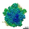 12332MC 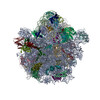 7nhkC 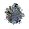 7nhmC 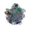 7nhnC M: map data used to model this data C: citing same article ( |
|---|---|
| Similar structure data | |
| EM raw data | EMPIAR-10683 (Title: Affinity-purified VgaA-LC in complex with 70S ribosomes from Staphylococcus aureus Data size: 241.0 Data #1: Unaligned multi-frame micrographs of VgaA-LC bound to 70S ribosome from Staphyloccous aureus [micrographs - multiframe]) |

-Links
 PDBj
PDBj


- Assembly
| Deposited unit | 
|
|---|---|
| 1 |
|
-Components
-Protein , 1 types, 1 molecules 0
| #1: Protein | Mass: 63265.316 Da / Num. of mol.: 1 Source method: isolated from a genetically manipulated source Source: (gene. exp.) Staphylococcus haemolyticus (bacteria) / Gene: vga(A)LCProduction host: Staphylococcus aureus subsp. aureus NCTC 8325 (bacteria)Strain (production host): SH1000 / References: UniProt: A0SNL9 |
|---|
-RNA chain , 5 types, 5 molecules DAaBb
| #2: RNA chain | Mass: 24506.611 Da / Num. of mol.: 1 / Source method: isolated from a natural source Source: (natural) Staphylococcus aureus subsp. aureus NCTC 8325 (bacteria) |
|---|---|
| #5: RNA chain | Mass: 946696.688 Da / Num. of mol.: 1 / Source method: isolated from a natural source Source: (natural) Staphylococcus aureus subsp. aureus NCTC 8325 (bacteria)References: GenBank: 87201381 |
| #6: RNA chain | Mass: 501711.156 Da / Num. of mol.: 1 / Source method: isolated from a natural source Source: (natural) Staphylococcus aureus subsp. aureus NCTC 8325 (bacteria)References: GenBank: CP000253.1 |
| #7: RNA chain | Mass: 36974.945 Da / Num. of mol.: 1 / Source method: isolated from a natural source Source: (natural) Staphylococcus aureus subsp. aureus NCTC 8325 (bacteria)References: GenBank: CP000253.1 |
| #48: RNA chain | Mass: 4128.384 Da / Num. of mol.: 1 / Source method: isolated from a natural source Source: (natural) Staphylococcus aureus subsp. aureus NCTC 8325 (bacteria) |
+50S ribosomal protein ... , 28 types, 28 molecules ZPGHIJKMNOQRSTUVWXY123687945
-30S ribosomal protein ... , 19 types, 19 molecules dehklopqutnjfisgmrc
| #31: Protein | Mass: 24143.867 Da / Num. of mol.: 1 / Source method: isolated from a natural source Source: (natural) Staphylococcus aureus subsp. aureus NCTC 8325 (bacteria)References: UniProt: Q2FW12 |
|---|---|
| #32: Protein | Mass: 23051.416 Da / Num. of mol.: 1 / Source method: isolated from a natural source Source: (natural) Staphylococcus aureus subsp. aureus NCTC 8325 (bacteria)References: UniProt: Q2FXK6 |
| #33: Protein | Mass: 17826.555 Da / Num. of mol.: 1 / Source method: isolated from a natural source Source: (natural) Staphylococcus aureus subsp. aureus NCTC 8325 (bacteria)References: UniProt: P48940 |
| #34: Protein | Mass: 11598.503 Da / Num. of mol.: 1 / Source method: isolated from a natural source Source: (natural) Staphylococcus aureus subsp. aureus NCTC 8325 (bacteria)References: UniProt: A0A0D1JTN2 |
| #35: Protein | Mass: 13907.978 Da / Num. of mol.: 1 / Source method: isolated from a natural source Source: (natural) Staphylococcus aureus subsp. aureus NCTC 8325 (bacteria)References: UniProt: Q2FW31 |
| #36: Protein | Mass: 7246.691 Da / Num. of mol.: 1 / Source method: isolated from a natural source Source: (natural) Staphylococcus aureus subsp. aureus NCTC 8325 (bacteria)References: UniProt: Q2FW19 |
| #37: Protein | Mass: 10634.330 Da / Num. of mol.: 1 / Source method: isolated from a natural source Source: (natural) Staphylococcus aureus subsp. aureus NCTC 8325 (bacteria)References: UniProt: Q2G2Q1 |
| #38: Protein | Mass: 10253.886 Da / Num. of mol.: 1 / Source method: isolated from a natural source Source: (natural) Staphylococcus aureus subsp. aureus NCTC 8325 (bacteria)References: UniProt: Q2FZ45 |
| #39: Protein | Mass: 9039.472 Da / Num. of mol.: 1 / Source method: isolated from a natural source Source: (natural) Staphylococcus aureus subsp. aureus NCTC 8325 (bacteria)References: UniProt: Q2FXY6 |
| #41: Protein | Mass: 10639.309 Da / Num. of mol.: 1 / Source method: isolated from a natural source Source: (natural) Staphylococcus aureus subsp. aureus NCTC 8325 (bacteria)References: UniProt: Q2FW10 |
| #42: Protein | Mass: 13747.919 Da / Num. of mol.: 1 / Source method: isolated from a natural source Source: (natural) Staphylococcus aureus subsp. aureus NCTC 8325 (bacteria)References: UniProt: Q2FW30 |
| #43: Protein | Mass: 14856.987 Da / Num. of mol.: 1 / Source method: isolated from a natural source Source: (natural) Staphylococcus aureus subsp. aureus NCTC 8325 (bacteria)References: UniProt: Q2FW39 |
| #44: Protein | Mass: 17770.512 Da / Num. of mol.: 1 / Source method: isolated from a natural source Source: (natural) Staphylococcus aureus subsp. aureus NCTC 8325 (bacteria)References: UniProt: Q2FW23 |
| #45: Protein | Mass: 14854.315 Da / Num. of mol.: 1 / Source method: isolated from a natural source Source: (natural) Staphylococcus aureus subsp. aureus NCTC 8325 (bacteria)References: UniProt: Q2FW20 |
| #46: Protein | Mass: 9332.018 Da / Num. of mol.: 1 / Source method: isolated from a natural source Source: (natural) Staphylococcus aureus subsp. aureus NCTC 8325 (bacteria)References: UniProt: Q2G111 |
| #47: Protein | Mass: 11613.146 Da / Num. of mol.: 1 / Source method: isolated from a natural source Source: (natural) Staphylococcus aureus subsp. aureus NCTC 8325 (bacteria)References: UniProt: Q2G113 |
| #49: Protein | Mass: 15320.870 Da / Num. of mol.: 1 / Source method: isolated from a natural source Source: (natural) Staphylococcus aureus subsp. aureus NCTC 8325 (bacteria)References: UniProt: P0A0H0 |
| #50: Protein | Mass: 10196.888 Da / Num. of mol.: 1 / Source method: isolated from a natural source Source: (natural) Staphylococcus aureus subsp. aureus NCTC 8325 (bacteria)References: UniProt: Q2FW15 |
| #51: Protein | Mass: 29136.369 Da / Num. of mol.: 1 / Source method: isolated from a natural source Source: (natural) Staphylococcus aureus subsp. aureus NCTC 8325 (bacteria)References: UniProt: Q2FZ25 |
-Non-polymers , 4 types, 162 molecules 


| #54: Chemical | ChemComp-MG /  Mass: 24.305 Da / Num. of mol.: 138 / Source method: obtained synthetically / Formula: Mg Mass: 24.305 Da / Num. of mol.: 138 / Source method: obtained synthetically / Formula: Mg#55: Chemical |  Mass: 507.181 Da / Num. of mol.: 2 / Source method: obtained synthetically / Formula: C10H16N5O13P3 / Comment: ATP, energy-carrying molecule*YM Mass: 507.181 Da / Num. of mol.: 2 / Source method: obtained synthetically / Formula: C10H16N5O13P3 / Comment: ATP, energy-carrying molecule*YM#56: Chemical | ChemComp-K /  Mass: 39.098 Da / Num. of mol.: 18 / Source method: obtained synthetically / Formula: K Mass: 39.098 Da / Num. of mol.: 18 / Source method: obtained synthetically / Formula: K#57: Chemical | ChemComp-ZN /  Mass: 65.409 Da / Num. of mol.: 4 / Source method: obtained synthetically / Formula: Zn Mass: 65.409 Da / Num. of mol.: 4 / Source method: obtained synthetically / Formula: Zn |
|---|
-Details
| Has ligand of interest | N |
|---|
-Experimental details
-Experiment
| Experiment | Method: ELECTRON MICROSCOPY |
|---|---|
| EM experiment | Aggregation state: PARTICLE / 3D reconstruction method: single particle reconstruction |
- Sample preparation
| Component |
| ||||||||||||||||||||||||
|---|---|---|---|---|---|---|---|---|---|---|---|---|---|---|---|---|---|---|---|---|---|---|---|---|---|
| Molecular weight | Value: 2.2 MDa / Experimental value: NO | ||||||||||||||||||||||||
| Source (natural) |
| ||||||||||||||||||||||||
| Source (recombinant) | Organism: Staphylococcus aureus subsp. aureus NCTC 8325 (bacteria) Strain: SH1000 | ||||||||||||||||||||||||
| Buffer solution | pH: 7.5 | ||||||||||||||||||||||||
| Specimen | Embedding applied: NO / Shadowing applied: NO / Staining applied: NO / Vitrification applied: YES | ||||||||||||||||||||||||
| Specimen support | Grid material: COPPER / Grid mesh size: 200 divisions/in. / Grid type: Quantifoil R1.2/1.3 | ||||||||||||||||||||||||
| Vitrification | Instrument: FEI VITROBOT MARK III / Cryogen name: ETHANE / Humidity: 100 % / Chamber temperature: 277.15 K |
- Electron microscopy imaging
| Experimental equipment |  Model: Titan Krios / Image courtesy: FEI Company |
|---|---|
| Microscopy | Model: FEI TITAN KRIOS |
| Electron gun | Electron source:  FIELD EMISSION GUN / Accelerating voltage: 300 kV / Illumination mode: FLOOD BEAM FIELD EMISSION GUN / Accelerating voltage: 300 kV / Illumination mode: FLOOD BEAM |
| Electron lens | Mode: BRIGHT FIELD / Nominal magnification: 165000 X / Nominal defocus max: -1900 nm / Nominal defocus min: -700 nm / Cs: 2.7 mm |
| Image recording | Electron dose: 26.3 e/Å2 / Film or detector model: GATAN K2 SUMMIT (4k x 4k) / Num. of grids imaged: 1 |
- Processing
| EM software | Name: Gctf / Category: CTF correction |
|---|---|
| CTF correction | Type: PHASE FLIPPING AND AMPLITUDE CORRECTION |
| Particle selection | Num. of particles selected: 165872 |
| 3D reconstruction | Resolution: 3.1 Å / Resolution method: FSC 0.143 CUT-OFF / Num. of particles: 61910 / Num. of class averages: 1 / Symmetry type: POINT |
