 Movie
Movie Controller

[English] 日本語
 Yorodumi
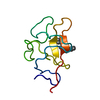
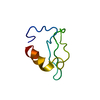
- PDB-4omo: Crystal structure of the c-Src tyrosine kinase SH3 domain mutant Q128E -
+ Open data

- Basic information
| Entry | Database: PDB / ID: 4omo | ||||||
|---|---|---|---|---|---|---|---|
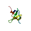
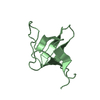
| Title | Crystal structure of the c-Src tyrosine kinase SH3 domain mutant Q128E | ||||||
 Components Components | Proto-oncogene tyrosine-protein kinase Src | ||||||
 Keywords Keywords | TRANSFERASE / beta-barrel sandwich / kinase / proline rich motifs | ||||||
| Function / homology |  Function and homology information Function and homology informationSignaling by ERBB2 / Nuclear signaling by ERBB4 / Signaling by SCF-KIT / Regulation of KIT signaling / Signaling by EGFR / GAB1 signalosome / Regulation of gap junction activity / FCGR activation / PECAM1 interactions / Co-stimulation by CD28 ...Signaling by ERBB2 / Nuclear signaling by ERBB4 / Signaling by SCF-KIT / Regulation of KIT signaling / Signaling by EGFR / GAB1 signalosome / Regulation of gap junction activity / FCGR activation / PECAM1 interactions / Co-stimulation by CD28 / Co-inhibition by CTLA4 / EPHA-mediated growth cone collapse / Ephrin signaling / G alpha (i) signalling events / GP1b-IX-V activation signalling / Thrombin signalling through proteinase activated receptors (PARs) / VEGFR2 mediated cell proliferation / RET signaling / Receptor Mediated Mitophagy / : / ADP signalling through P2Y purinoceptor 1 / RAF activation / PIP3 activates AKT signaling / EPH-ephrin mediated repulsion of cells / PI5P, PP2A and IER3 Regulate PI3K/AKT Signaling / Downstream signal transduction / Downregulation of ERBB4 signaling / Cyclin D associated events in G1 / Regulation of RUNX3 expression and activity / Degradation of CDH1 / MAP2K and MAPK activation / Integrin signaling / GRB2:SOS provides linkage to MAPK signaling for Integrins / : / MET activates PTK2 signaling / Extra-nuclear estrogen signaling / EPHB-mediated forward signaling / p130Cas linkage to MAPK signaling for integrins / VEGFA-VEGFR2 Pathway / connexin binding / negative regulation of intrinsic apoptotic signaling pathway / progesterone receptor signaling pathway / immune system process / negative regulation of extrinsic apoptotic signaling pathway / non-specific protein-tyrosine kinase / non-membrane spanning protein tyrosine kinase activity / epidermal growth factor receptor signaling pathway / cell junction / cell-cell junction / protein tyrosine kinase activity / protein phosphatase binding / cytoskeleton / cell differentiation / cell adhesion / regulation of cell cycle / endosome membrane / mitochondrial inner membrane / signaling receptor binding / focal adhesion / heme binding / perinuclear region of cytoplasm / protein-containing complex / ATP binding / membrane / nucleus / plasma membrane / cytosol Similarity search - Function | ||||||
| Biological species |  | ||||||
| Method |  X-RAY DIFFRACTION / SYNCHROTRON / MOLECULAR REPLACEMENT / molecular replacement / Resolution: 1.04 Å X-RAY DIFFRACTION / SYNCHROTRON / MOLECULAR REPLACEMENT / molecular replacement / Resolution: 1.04 Å | ||||||
 Authors Authors | Camara-Artigas, A. / Bacarizo, J. | ||||||
 Citation Citation | Journal: Plos One / Year: 2014 Title: Electrostatic Effects in the Folding of the SH3 Domain of the c-Src Tyrosine Kinase: pH-Dependence in 3D-Domain Swapping and Amyloid Formation. Authors: Bacarizo, J. / Martinez-Rodriguez, S. / Martin-Garcia, J.M. / Andujar-Sanchez, M. / Ortiz-Salmeron, E. / Neira, J.L. / Camara-Artigas, A. | ||||||
| History |
|
- Structure visualization
| Structure viewer | Molecule: MolmilJmol/JSmol |
|---|
- Downloads & links
-Download
| PDBx/mmCIF format | 4omo.cif.gz | 93.3 KB | Display | PDBx/mmCIF format |
|---|---|---|---|---|
| PDB format | pdb4omo.ent.gz | 71.3 KB | Display | PDB format |
| PDBx/mmJSON format | 4omo.json.gz | Tree view | PDBx/mmJSON format | |
| Others |  Other downloads Other downloads |
-Validation report
| Arichive directory | https://data.pdbj.org/pub/pdb/validation_reports/om/4omoftp://data.pdbj.org/pub/pdb/validation_reports/om/4omo | HTTPS FTP |
|---|
-Related structure data
| Related structure data |  4jz3C  4jz4SC  4omlC  4omnC  4ompC C: citing same article ( S: Starting model for refinement |
|---|---|
| Similar structure data |


-Links
 PDBj
PDBj- Assembly
| Deposited unit | 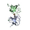
| ||||||||
|---|---|---|---|---|---|---|---|---|---|
| 1 | 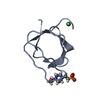
| ||||||||
| 2 | 
| ||||||||
| Unit cell |
|
-Components
| #1: Protein | Mass: 6906.483 Da / Num. of mol.: 2 / Fragment: SH3 domain / Mutation: Q128E Source method: isolated from a genetically manipulated source Source: (gene. exp.)  References: UniProt: P00523, non-specific protein-tyrosine kinase #2: Chemical |   Mass: 58.693 Da / Num. of mol.: 2 / Source method: obtained synthetically / Formula: Ni Mass: 58.693 Da / Num. of mol.: 2 / Source method: obtained synthetically / Formula: Ni#3: Chemical | ChemComp-EPE / |   Mass: 238.305 Da / Num. of mol.: 1 / Source method: obtained synthetically / Formula: C8H18N2O4S / Comment: pH buffer*YM Mass: 238.305 Da / Num. of mol.: 1 / Source method: obtained synthetically / Formula: C8H18N2O4S / Comment: pH buffer*YM#4: Water | ChemComp-HOH / |  Mass: 18.015 Da / Num. of mol.: 155 / Source method: isolated from a natural source / Formula: H2O Mass: 18.015 Da / Num. of mol.: 155 / Source method: isolated from a natural source / Formula: H2O |
|---|
-Experimental details
-Experiment
| Experiment | Method: X-RAY DIFFRACTION / Number of used crystals: 1 |
|---|
- Sample preparation
| Crystal | Density Matthews: 2.08 Å3/Da / Density % sol: 40.84 % |
|---|---|
| Crystal grow | Temperature: 298 K / Method: vapor diffusion, hanging drop / pH: 7.5 Details: 0.1M Hepes, 2M ammonium sulphate, 5mM NiCl2, 5mM MBCD, pH 7.5, VAPOR DIFFUSION, HANGING DROP, temperature 298K |
-Data collection
| Diffraction | Mean temperature: 100 K | |||||||||||||||||||||
|---|---|---|---|---|---|---|---|---|---|---|---|---|---|---|---|---|---|---|---|---|---|---|
| Diffraction source | Source: SYNCHROTRON / Site: ALBA  / Beamline: XALOC / Wavelength: 1.033198 Å / Beamline: XALOC / Wavelength: 1.033198 Å | |||||||||||||||||||||
| Detector | Type: DECTRIS PILATUS 6M / Detector: PIXEL / Date: Jul 7, 2013 / Details: Channel-cut Si(111) + KB focusing mirrors | |||||||||||||||||||||
| Radiation | Monochromator: Si(111) channel-cut, cryocooled / Protocol: SINGLE WAVELENGTH / Monochromatic (M) / Laue (L): M / Scattering type: x-ray | |||||||||||||||||||||
| Radiation wavelength | Wavelength: 1.033198 Å / Relative weight: 1 | |||||||||||||||||||||
| Reflection | Redundancy: 12.1 % / Number: 684773 / Rmerge(I) obs: 0.069 / D res high: 1.04 Å / D res low: 45.46 Å / Num. obs: 56398 / % possible obs: 99.2 | |||||||||||||||||||||
| Diffraction reflection shell |
| |||||||||||||||||||||
| Reflection | Resolution: 1.04→45.46 Å / Num. all: 56853 / Num. obs: 53404 / % possible obs: 99.2 % / Observed criterion σ(F): 0 / Observed criterion σ(I): 0 / Redundancy: 12.1 % / Biso Wilson estimate: 10.57 Å2 / Rmerge(I) obs: 0.069 / Rsym value: 0.069 / Net I/σ(I): 20.3 / Scaling rejects: 6 | |||||||||||||||||||||
| Reflection shell | Diffraction-ID: 1
|
-Phasing
| Phasing | Method: molecular replacement |
|---|
- Processing
| Software |
| ||||||||||||||||||||||||||||||||||||||||||||||||||||||||||||||||||||||||||||||||||||||||||||||||||||||||||||||||||||||||||||||||||||||||||||||||||||||||||||||||||||||||||||||||||||||||||||||||||||||||||||||||||||||||||||||||||||||||||||||||||||||||
|---|---|---|---|---|---|---|---|---|---|---|---|---|---|---|---|---|---|---|---|---|---|---|---|---|---|---|---|---|---|---|---|---|---|---|---|---|---|---|---|---|---|---|---|---|---|---|---|---|---|---|---|---|---|---|---|---|---|---|---|---|---|---|---|---|---|---|---|---|---|---|---|---|---|---|---|---|---|---|---|---|---|---|---|---|---|---|---|---|---|---|---|---|---|---|---|---|---|---|---|---|---|---|---|---|---|---|---|---|---|---|---|---|---|---|---|---|---|---|---|---|---|---|---|---|---|---|---|---|---|---|---|---|---|---|---|---|---|---|---|---|---|---|---|---|---|---|---|---|---|---|---|---|---|---|---|---|---|---|---|---|---|---|---|---|---|---|---|---|---|---|---|---|---|---|---|---|---|---|---|---|---|---|---|---|---|---|---|---|---|---|---|---|---|---|---|---|---|---|---|---|---|---|---|---|---|---|---|---|---|---|---|---|---|---|---|---|---|---|---|---|---|---|---|---|---|---|---|---|---|---|---|---|---|---|---|---|---|---|---|---|---|---|---|---|---|---|---|---|---|
| Refinement | Method to determine structure: MOLECULAR REPLACEMENT Starting model: pdb entry 4JZ4 Resolution: 1.04→24.109 Å / SU ML: 0.07 / Isotropic thermal model: anisotropic / Cross valid method: THROUGHOUT / σ(F): 0.27 / σ(I): 0 / Phase error: 15.56 / Stereochemistry target values: ML
| ||||||||||||||||||||||||||||||||||||||||||||||||||||||||||||||||||||||||||||||||||||||||||||||||||||||||||||||||||||||||||||||||||||||||||||||||||||||||||||||||||||||||||||||||||||||||||||||||||||||||||||||||||||||||||||||||||||||||||||||||||||||||
| Solvent computation | Shrinkage radii: 0.9 Å / VDW probe radii: 1.11 Å / Solvent model: FLAT BULK SOLVENT MODEL | ||||||||||||||||||||||||||||||||||||||||||||||||||||||||||||||||||||||||||||||||||||||||||||||||||||||||||||||||||||||||||||||||||||||||||||||||||||||||||||||||||||||||||||||||||||||||||||||||||||||||||||||||||||||||||||||||||||||||||||||||||||||||
| Displacement parameters | Biso max: 46.23 Å2 / Biso mean: 16.6867 Å2 / Biso min: 1.07 Å2 | ||||||||||||||||||||||||||||||||||||||||||||||||||||||||||||||||||||||||||||||||||||||||||||||||||||||||||||||||||||||||||||||||||||||||||||||||||||||||||||||||||||||||||||||||||||||||||||||||||||||||||||||||||||||||||||||||||||||||||||||||||||||||
| Refinement step | Cycle: LAST / Resolution: 1.04→24.109 Å
| ||||||||||||||||||||||||||||||||||||||||||||||||||||||||||||||||||||||||||||||||||||||||||||||||||||||||||||||||||||||||||||||||||||||||||||||||||||||||||||||||||||||||||||||||||||||||||||||||||||||||||||||||||||||||||||||||||||||||||||||||||||||||
| Refine LS restraints |
| ||||||||||||||||||||||||||||||||||||||||||||||||||||||||||||||||||||||||||||||||||||||||||||||||||||||||||||||||||||||||||||||||||||||||||||||||||||||||||||||||||||||||||||||||||||||||||||||||||||||||||||||||||||||||||||||||||||||||||||||||||||||||
| LS refinement shell | Refine-ID: X-RAY DIFFRACTION / Total num. of bins used: 30
|
