 Movie
Movie Controller

[English] 日本語
 Yorodumi
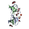
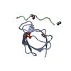
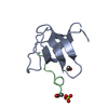
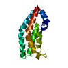
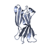
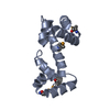
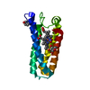
- PDB-4oml: Crystal structure of the intertwined dimer of the c-Src tyrosine ... -
+ Open data

- Basic information
| Entry | Database: PDB / ID: 4oml | ||||||
|---|---|---|---|---|---|---|---|
| Title | Crystal structure of the intertwined dimer of the c-Src tyrosine kinase SH3 domain mutant Q128R | ||||||
 Components Components | Proto-oncogene tyrosine-protein kinase Src | ||||||
 Keywords Keywords | TRANSFERASE / beta-barrel sandwich / kinase / proline rich motifs | ||||||
| Function / homology |  Function and homology information Function and homology informationSignaling by ERBB2 / Nuclear signaling by ERBB4 / Signaling by SCF-KIT / Regulation of KIT signaling / Signaling by EGFR / GAB1 signalosome / Regulation of gap junction activity / FCGR activation / PECAM1 interactions / Co-stimulation by CD28 ...Signaling by ERBB2 / Nuclear signaling by ERBB4 / Signaling by SCF-KIT / Regulation of KIT signaling / Signaling by EGFR / GAB1 signalosome / Regulation of gap junction activity / FCGR activation / PECAM1 interactions / Co-stimulation by CD28 / Co-inhibition by CTLA4 / EPHA-mediated growth cone collapse / Ephrin signaling / G alpha (i) signalling events / GP1b-IX-V activation signalling / Thrombin signalling through proteinase activated receptors (PARs) / VEGFR2 mediated cell proliferation / RET signaling / Receptor Mediated Mitophagy / : / ADP signalling through P2Y purinoceptor 1 / RAF activation / PIP3 activates AKT signaling / EPH-ephrin mediated repulsion of cells / PI5P, PP2A and IER3 Regulate PI3K/AKT Signaling / Downstream signal transduction / Downregulation of ERBB4 signaling / Cyclin D associated events in G1 / Regulation of RUNX3 expression and activity / Degradation of CDH1 / MAP2K and MAPK activation / Integrin signaling / GRB2:SOS provides linkage to MAPK signaling for Integrins / : / MET activates PTK2 signaling / Extra-nuclear estrogen signaling / EPHB-mediated forward signaling / p130Cas linkage to MAPK signaling for integrins / VEGFA-VEGFR2 Pathway / connexin binding / negative regulation of intrinsic apoptotic signaling pathway / progesterone receptor signaling pathway / immune system process / negative regulation of extrinsic apoptotic signaling pathway / non-specific protein-tyrosine kinase / non-membrane spanning protein tyrosine kinase activity / epidermal growth factor receptor signaling pathway / cell junction / cell-cell junction / protein tyrosine kinase activity / protein phosphatase binding / cytoskeleton / cell differentiation / cell adhesion / regulation of cell cycle / endosome membrane / mitochondrial inner membrane / signaling receptor binding / focal adhesion / heme binding / perinuclear region of cytoplasm / protein-containing complex / ATP binding / membrane / nucleus / plasma membrane / cytosol Similarity search - Function | ||||||
| Biological species |  | ||||||
| Method |  X-RAY DIFFRACTION / SYNCHROTRON / MOLECULAR REPLACEMENT / molecular replacement / Resolution: 1.6 Å X-RAY DIFFRACTION / SYNCHROTRON / MOLECULAR REPLACEMENT / molecular replacement / Resolution: 1.6 Å | ||||||
 Authors Authors | Camara-Artigas, A. / Bacarizo, J. | ||||||
 Citation Citation | Journal: Plos One / Year: 2014 Title: Electrostatic Effects in the Folding of the SH3 Domain of the c-Src Tyrosine Kinase: pH-Dependence in 3D-Domain Swapping and Amyloid Formation. Authors: Bacarizo, J. / Martinez-Rodriguez, S. / Martin-Garcia, J.M. / Andujar-Sanchez, M. / Ortiz-Salmeron, E. / Neira, J.L. / Camara-Artigas, A. | ||||||
| History |
|
- Structure visualization
| Structure viewer | Molecule: MolmilJmol/JSmol |
|---|
- Downloads & links
-Download
| PDBx/mmCIF format | 4oml.cif.gz | 51.3 KB | Display | PDBx/mmCIF format |
|---|---|---|---|---|
| PDB format | pdb4oml.ent.gz | 36.2 KB | Display | PDB format |
| PDBx/mmJSON format | 4oml.json.gz | Tree view | PDBx/mmJSON format | |
| Others |  Other downloads Other downloads |
-Validation report
| Arichive directory | https://data.pdbj.org/pub/pdb/validation_reports/om/4omlftp://data.pdbj.org/pub/pdb/validation_reports/om/4oml | HTTPS FTP |
|---|
-Related structure data
| Related structure data | 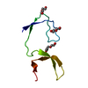 4jz3C 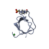 4jz4SC 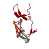 4omnC 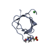 4omoC 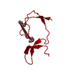 4ompC C: citing same article ( S: Starting model for refinement |
|---|---|
| Similar structure data |


-Links
 PDBj
PDBj- Assembly
| Deposited unit | 
| ||||||||
|---|---|---|---|---|---|---|---|---|---|
| 1 | 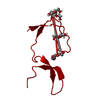
| ||||||||
| Unit cell |
| ||||||||
| Components on special symmetry positions |
| ||||||||
| Details | Intertwined dimer. The dimer is generated by symmetry |
-Components
| #1: Protein | Mass: 8823.652 Da / Num. of mol.: 1 / Fragment: SH3 domain / Mutation: Q128R Source method: isolated from a genetically manipulated source Source: (gene. exp.)  References: UniProt: P00523, non-specific protein-tyrosine kinase | ||
|---|---|---|---|
| #2: Chemical | ChemComp-PGE /   Mass: 150.173 Da / Num. of mol.: 1 / Source method: obtained synthetically / Formula: C6H14O4 Mass: 150.173 Da / Num. of mol.: 1 / Source method: obtained synthetically / Formula: C6H14O4 | ||
| #3: Chemical |   Mass: 106.120 Da / Num. of mol.: 3 / Source method: obtained synthetically / Formula: C4H10O3 Mass: 106.120 Da / Num. of mol.: 3 / Source method: obtained synthetically / Formula: C4H10O3#4: Water | ChemComp-HOH / |  Mass: 18.015 Da / Num. of mol.: 22 / Source method: isolated from a natural source / Formula: H2O Mass: 18.015 Da / Num. of mol.: 22 / Source method: isolated from a natural source / Formula: H2O |
-Experimental details
-Experiment
| Experiment | Method: X-RAY DIFFRACTION / Number of used crystals: 1 |
|---|
- Sample preparation
| Crystal | Density Matthews: 2.27 Å3/Da / Density % sol: 45.79 % |
|---|---|
| Crystal grow | Temperature: 298 K / pH: 5 Details: 0.1M sodium acetate, 1.6 M ammonium sulphate, 10%PEG 300, 10%Glicerol, pH 5.0, vapor diffusion, hanging drop, temperature 298K |
-Data collection
| Diffraction | Mean temperature: 100 K | ||||||||||||||||||
|---|---|---|---|---|---|---|---|---|---|---|---|---|---|---|---|---|---|---|---|
| Diffraction source | Source: SYNCHROTRON / Site: ESRF  / Beamline: BM16 / Wavelength: 0.97752 / Beamline: BM16 / Wavelength: 0.97752 | ||||||||||||||||||
| Detector | Type: MAR CCD 165 mm / Detector: CCD / Date: Mar 3, 2008 / Details: MIRRORS | ||||||||||||||||||
| Radiation | Monochromator: SI(111) CHANNEL-CUT / Protocol: SINGLE WAVELENGTH / Monochromatic (M) / Laue (L): M / Scattering type: x-ray | ||||||||||||||||||
| Radiation wavelength | Wavelength: 0.97752 Å / Relative weight: 1 | ||||||||||||||||||
| Reflection | Redundancy: 12.9 % / Number: 143425 / Rmerge(I) obs: 0.073 / D res high: 1.6 Å / D res low: 19.22 Å / Num. obs: 11086 / % possible obs: 97 | ||||||||||||||||||
| Diffraction reflection shell |
| ||||||||||||||||||
| Reflection | Resolution: 1.6→19.22 Å / Num. obs: 11086 / % possible obs: 97 % / Observed criterion σ(I): 0 / Redundancy: 12.9 % / Biso Wilson estimate: 21.57 Å2 / Rmerge(I) obs: 0.073 / Rsym value: 0.073 / Net I/σ(I): 21.4 | ||||||||||||||||||
| Reflection shell | Resolution: 1.6→1.63 Å / Redundancy: 13 % / Rmerge(I) obs: 0.654 / Mean I/σ(I) obs: 4.4 / % possible all: 93.9 |
-Phasing
| Phasing | Method: molecular replacement |
|---|
- Processing
| Software |
| ||||||||||||||||||||||||||||||||||||||||||||||||||||||||
|---|---|---|---|---|---|---|---|---|---|---|---|---|---|---|---|---|---|---|---|---|---|---|---|---|---|---|---|---|---|---|---|---|---|---|---|---|---|---|---|---|---|---|---|---|---|---|---|---|---|---|---|---|---|---|---|---|---|
| Refinement | Method to determine structure: MOLECULAR REPLACEMENT Starting model: PDB ENTRY 4JZ4 Resolution: 1.6→19.22 Å / SU ML: 0.12 / Isotropic thermal model: anisotropic / σ(F): 1.45 / Phase error: 17.9 / Stereochemistry target values: ML
| ||||||||||||||||||||||||||||||||||||||||||||||||||||||||
| Solvent computation | Shrinkage radii: 0.9 Å / VDW probe radii: 1.11 Å / Solvent model: FLAT BULK SOLVENT MODEL | ||||||||||||||||||||||||||||||||||||||||||||||||||||||||
| Displacement parameters | Biso mean: 37.89 Å2 | ||||||||||||||||||||||||||||||||||||||||||||||||||||||||
| Refinement step | Cycle: LAST / Resolution: 1.6→19.22 Å
| ||||||||||||||||||||||||||||||||||||||||||||||||||||||||
| Refine LS restraints |
| ||||||||||||||||||||||||||||||||||||||||||||||||||||||||
| LS refinement shell |
|
