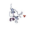
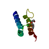
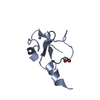
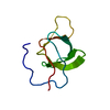
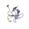
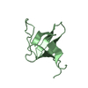
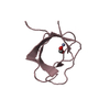
[English] 日本語
Yorodumi
- PDB-3ua7: Crystal Structure of the Human Fyn SH3 domain in complex with a p... -

+
Open data


ID or keywords:

Loading...

-
Basic information

Entry
Database: PDB / ID: 3ua7
TitleCrystal Structure of the Human Fyn SH3 domain in complex with a peptide from the Hepatitis C virus NS5A-protein
Components
  • Non-structural protein 5A
  • Tyrosine-protein kinase Fyn
KeywordsTRANSFERASE/VIRAL PROTEIN / beta barrel / kinase / poly proline rich motif / TRANSFERASE-VIRAL PROTEIN complex
Function / homology
Function and homology information


response to singlet oxygen / Reelin signalling pathway / negative regulation of hydrogen peroxide biosynthetic process / perinuclear endoplasmic reticulum / NTRK2 activates RAC1 / growth factor receptor binding / Activated NTRK2 signals through FYN / heart process / cellular response to L-glutamate / regulation of glutamate receptor signaling pathway ...response to singlet oxygen / Reelin signalling pathway / negative regulation of hydrogen peroxide biosynthetic process / perinuclear endoplasmic reticulum / NTRK2 activates RAC1 / growth factor receptor binding / Activated NTRK2 signals through FYN / heart process / cellular response to L-glutamate / regulation of glutamate receptor signaling pathway / regulation of calcium ion import across plasma membrane / SEMA3A-Plexin repulsion signaling by inhibiting Integrin adhesion / G protein-coupled glutamate receptor signaling pathway / Platelet Adhesion to exposed collagen / CD28 co-stimulation / positive regulation of protein localization to membrane / activated T cell proliferation / CRMPs in Sema3A signaling / positive regulation of cysteine-type endopeptidase activity / FLT3 signaling through SRC family kinases / negative regulation of dendritic spine maintenance / feeding behavior / Nef and signal transduction / type 5 metabotropic glutamate receptor binding / Nephrin family interactions / DCC mediated attractive signaling / dendrite morphogenesis / EPH-Ephrin signaling / CD28 dependent Vav1 pathway / hepacivirin / Ephrin signaling / dendritic spine maintenance / Regulation of KIT signaling / host cell mitochondrial membrane / tau-protein kinase activity / CTLA4 inhibitory signaling / host cell lipid droplet / leukocyte migration / phospholipase activator activity / Fc-gamma receptor signaling pathway involved in phagocytosis / EPHA-mediated growth cone collapse / symbiont-mediated suppression of host TRAF-mediated signal transduction / cellular response to platelet-derived growth factor stimulus / transformation of host cell by virus / Dectin-2 family / symbiont-mediated perturbation of host cell cycle G1/S transition checkpoint / stimulatory C-type lectin receptor signaling pathway / PECAM1 interactions / CD28 dependent PI3K/Akt signaling / phospholipase binding / response to amyloid-beta / glial cell projection / Sema3A PAK dependent Axon repulsion / cellular response to glycine / FCGR activation / alpha-tubulin binding / EPH-ephrin mediated repulsion of cells / positive regulation of protein targeting to membrane / ephrin receptor signaling pathway / Role of LAT2/NTAL/LAB on calcium mobilization / vascular endothelial growth factor receptor signaling pathway / detection of mechanical stimulus involved in sensory perception of pain / negative regulation of oxidative stress-induced intrinsic apoptotic signaling pathway / regulation of peptidyl-tyrosine phosphorylation / positive regulation of tyrosine phosphorylation of STAT protein / forebrain development / negative regulation of inflammatory response to antigenic stimulus / extrinsic component of cytoplasmic side of plasma membrane / Signaling by ERBB2 / negative regulation of protein ubiquitination / GPVI-mediated activation cascade / cellular response to transforming growth factor beta stimulus / cell surface receptor protein tyrosine kinase signaling pathway / symbiont-mediated suppression of host JAK-STAT cascade via inhibition of STAT1 activity / T cell costimulation / EPHB-mediated forward signaling / symbiont-mediated suppression of host cytoplasmic pattern recognition receptor signaling pathway via inhibition of MAVS activity / ephrin receptor binding / NCAM signaling for neurite out-growth / CD209 (DC-SIGN) signaling / ribonucleoside triphosphate phosphatase activity / FCGR3A-mediated IL10 synthesis / Signaling by phosphorylated juxtamembrane, extracellular and kinase domain KIT mutants / Antigen activates B Cell Receptor (BCR) leading to generation of second messengers / learning / actin filament / Regulation of signaling by CBL / Cell surface interactions at the vascular wall / FCGR3A-mediated phagocytosis / axon guidance / non-specific protein-tyrosine kinase / neuron migration / non-membrane spanning protein tyrosine kinase activity / Schaffer collateral - CA1 synapse / protein catabolic process / modulation of chemical synaptic transmission / tau protein binding / Signaling by SCF-KIT / negative regulation of protein catabolic process / VEGFA-VEGFR2 Pathway
Similarity search - Function
: / Fyn/Yrk, SH3 domain / : / Hepatitis C virus core protein, chain A superfamily / Hepatitus C virus, Non-structural 5a protein, C-terminal / Hepatitis C virus NS5A, 1B domain superfamily / Hepatitis C virus non-structural protein NS2, C-terminal domain / Hepatitis C virus non-structural protein NS2, N-terminal domain / Hepatitis C virus non-structural protein NS2 / HCV NS5a protein C-terminal region ...: / Fyn/Yrk, SH3 domain / : / Hepatitis C virus core protein, chain A superfamily / Hepatitus C virus, Non-structural 5a protein, C-terminal / Hepatitis C virus NS5A, 1B domain superfamily / Hepatitis C virus non-structural protein NS2, C-terminal domain / Hepatitis C virus non-structural protein NS2, N-terminal domain / Hepatitis C virus non-structural protein NS2 / HCV NS5a protein C-terminal region / Hepatitis C virus, Non-structural protein NS4b / Hepatitis C virus, Core protein, N-terminal / Hepatitis C virus non-structural protein NS4b / Hepatitis C virus capsid protein / Hepatitis C virus, Non-structural protein NS2 / Hepatitis C virus, Non-structural 5a protein / Hepatitis C virus, Non-structural 5a protein, domain 1a / Hepatitis C virus non-structural 5a, 1B domain / NS5A domain 1a superfamily / Hepatitis C virus non-structural 5a protein membrane anchor / Hepatitis C virus non-structural 5a zinc finger domain / Hepatitis C virus non-structural 5a domain 1b / Hepacivirus nonstructural protein 2 (NS2) protease domain profile. / Hepatitis C virus, Non-structural protein NS4a / Hepatitis C virus non-structural protein NS4a / Hepatitis C virus, Core protein, C-terminal / Hepatitis C virus core protein / Hepatitis C virus, Non-structural protein E2/NS1 / Hepatitis C virus non-structural protein E2/NS1 / Hepatitis C virus, Envelope glycoprotein E1 / Hepatitis C virus envelope glycoprotein E1 / RNA dependent RNA polymerase, hepatitis C virus / Viral RNA dependent RNA polymerase / Hepatitis C virus, NS3 protease, Peptidase S29 / Hepatitis C virus NS3 protease / Hepacivirus/Pegivirus NS3 protease domain profile. / SH3 Domains / DEAD box, Flavivirus / Flavivirus DEAD domain / SH3 domain / SH2 domain / Src homology 2 (SH2) domain profile. / Src homology 2 domains / SH3 type barrels. / SH2 domain / Src homology 3 domains / SH2 domain superfamily / SH3-like domain superfamily / Src homology 3 (SH3) domain profile. / SH3 domain / Tyrosine-protein kinase, catalytic domain / Tyrosine kinase, catalytic domain / Tyrosine protein kinases specific active-site signature. / Superfamilies 1 and 2 helicase C-terminal domain profile. / Superfamilies 1 and 2 helicase ATP-binding type-1 domain profile. / DEAD-like helicases superfamily / Helicase, C-terminal / Helicase superfamily 1/2, ATP-binding domain / Tyrosine-protein kinase, active site / Protein tyrosine and serine/threonine kinase / Serine-threonine/tyrosine-protein kinase, catalytic domain / Roll / Reverse transcriptase/Diguanylate cyclase domain / RNA-directed RNA polymerase, catalytic domain / RdRp of positive ssRNA viruses catalytic domain profile. / Peptidase S1, PA clan, chymotrypsin-like fold / Protein kinase, ATP binding site / Protein kinases ATP-binding region signature. / Peptidase S1, PA clan / DNA/RNA polymerase superfamily / Protein kinase domain profile. / Protein kinase domain / Protein kinase-like domain superfamily / P-loop containing nucleoside triphosphate hydrolase / Mainly Beta
Similarity search - Domain/homology
FORMIC ACID / Tyrosine-protein kinase Fyn / Genome polyprotein / Non-structural protein 5A
Similarity search - Component
Biological speciesHomo sapiens (human)
Hepatitis C virus
MethodX-RAY DIFFRACTION / SYNCHROTRON / MOLECULAR REPLACEMENT / molecular replacement / Resolution: 1.5 Å
AuthorsMartin-Garcia, J.M. / Ruiz-Sanz, J. / Luque, I. / Camara-Artigas, A.
CitationJournal: Acta Crystallogr.,Sect.D / Year: 2012
Title: The promiscuous binding of the Fyn SH3 domain to a peptide from the NS5A protein.
Authors: Martin-Garcia, J.M. / Luque, I. / Ruiz-Sanz, J. / Camara-Artigas, A.
History
DepositionOct 21, 2011Deposition site: RCSB / Processing site: RCSB
Revision 1.0Jul 25, 2012Provider: repository / Type: Initial release
Revision 1.1Jan 23, 2013Group: Database references
Revision 1.2Sep 13, 2023Group: Data collection / Database references ...Data collection / Database references / Derived calculations / Refinement description
Category: chem_comp_atom / chem_comp_bond ...chem_comp_atom / chem_comp_bond / database_2 / pdbx_initial_refinement_model / pdbx_struct_conn_angle / struct_conn / struct_ncs_dom_lim / struct_ref_seq_dif / struct_site
Item: _database_2.pdbx_DOI / _database_2.pdbx_database_accession ..._database_2.pdbx_DOI / _database_2.pdbx_database_accession / _pdbx_struct_conn_angle.ptnr1_auth_asym_id / _pdbx_struct_conn_angle.ptnr1_auth_comp_id / _pdbx_struct_conn_angle.ptnr1_auth_seq_id / _pdbx_struct_conn_angle.ptnr1_label_asym_id / _pdbx_struct_conn_angle.ptnr1_label_atom_id / _pdbx_struct_conn_angle.ptnr1_label_comp_id / _pdbx_struct_conn_angle.ptnr1_label_seq_id / _pdbx_struct_conn_angle.ptnr2_auth_asym_id / _pdbx_struct_conn_angle.ptnr2_auth_comp_id / _pdbx_struct_conn_angle.ptnr2_auth_seq_id / _pdbx_struct_conn_angle.ptnr2_label_asym_id / _pdbx_struct_conn_angle.ptnr2_label_atom_id / _pdbx_struct_conn_angle.ptnr2_label_comp_id / _pdbx_struct_conn_angle.ptnr3_auth_asym_id / _pdbx_struct_conn_angle.ptnr3_auth_comp_id / _pdbx_struct_conn_angle.ptnr3_auth_seq_id / _pdbx_struct_conn_angle.ptnr3_label_asym_id / _pdbx_struct_conn_angle.ptnr3_label_atom_id / _pdbx_struct_conn_angle.ptnr3_label_comp_id / _pdbx_struct_conn_angle.ptnr3_label_seq_id / _pdbx_struct_conn_angle.value / _struct_conn.pdbx_dist_value / _struct_conn.pdbx_leaving_atom_flag / _struct_conn.ptnr1_auth_asym_id / _struct_conn.ptnr1_auth_comp_id / _struct_conn.ptnr1_auth_seq_id / _struct_conn.ptnr1_label_asym_id / _struct_conn.ptnr1_label_atom_id / _struct_conn.ptnr1_label_comp_id / _struct_conn.ptnr1_label_seq_id / _struct_conn.ptnr2_auth_asym_id / _struct_conn.ptnr2_auth_comp_id / _struct_conn.ptnr2_auth_seq_id / _struct_conn.ptnr2_label_asym_id / _struct_conn.ptnr2_label_atom_id / _struct_conn.ptnr2_label_comp_id / _struct_conn.ptnr2_label_seq_id / _struct_ncs_dom_lim.beg_auth_comp_id / _struct_ncs_dom_lim.beg_label_asym_id / _struct_ncs_dom_lim.beg_label_comp_id / _struct_ncs_dom_lim.beg_label_seq_id / _struct_ncs_dom_lim.end_auth_comp_id / _struct_ncs_dom_lim.end_label_asym_id / _struct_ncs_dom_lim.end_label_comp_id / _struct_ncs_dom_lim.end_label_seq_id / _struct_ref_seq_dif.details / _struct_site.pdbx_auth_asym_id / _struct_site.pdbx_auth_comp_id / _struct_site.pdbx_auth_seq_id

-
Structure visualization

Structure viewerMolecule:
MolmilJmol/JSmol

Downloads & links

-
Assembly

Deposited unit
A: Tyrosine-protein kinase Fyn
B: Tyrosine-protein kinase Fyn
C: Tyrosine-protein kinase Fyn
D: Tyrosine-protein kinase Fyn
E: Non-structural protein 5A
F: Non-structural protein 5A
hetero molecules


Theoretical massNumber of molelcules
Total (without water)31,69013
Polymers31,3166
Non-polymers3737
Water3,117173
1
A: Tyrosine-protein kinase Fyn
E: Non-structural protein 5A
hetero molecules


Theoretical massNumber of molelcules
Total (without water)8,6375
Polymers8,4862
Non-polymers1513
Water362
TypeNameSymmetry operationNumber
identity operation1_555x,y,z1
2
D: Tyrosine-protein kinase Fyn
F: Non-structural protein 5A
hetero molecules


Theoretical massNumber of molelcules
Total (without water)8,5984
Polymers8,4862
Non-polymers1112
Water362
TypeNameSymmetry operationNumber
identity operation1_555x,y,z1
3
B: Tyrosine-protein kinase Fyn


Theoretical massNumber of molelcules
Total (without water)7,1721
Polymers7,1721
Non-polymers00
Water181
TypeNameSymmetry operationNumber
identity operation1_555x,y,z1
4
C: Tyrosine-protein kinase Fyn
hetero molecules


Theoretical massNumber of molelcules
Total (without water)7,2833
Polymers7,1721
Non-polymers1112
Water181
TypeNameSymmetry operationNumber
identity operation1_555x,y,z1
Unit cell
Length a, b, c (Å)51.390, 51.390, 185.590
Angle α, β, γ (deg.)90.000, 90.000, 90.000
Int Tables number92
Space group name H-MP41212
Components on special symmetry positions
IDModelComponents
11F-40-

HOH

Noncrystallographic symmetry (NCS)NCS domain:
IDEns-IDDetails
11A
21B
31C
41D

NCS domain segments:

Ens-ID: 1

Dom-IDComponent-IDBeg auth comp-IDBeg label comp-IDEnd auth comp-IDEnd label comp-IDRefine codeAuth asym-IDLabel asym-IDAuth seq-IDLabel seq-ID
11THRTHRSERSER2AA85 - 1146 - 35
21THRTHRSERSER2BB85 - 1146 - 35
31THRTHRSERSER2CC85 - 1146 - 35
41THRTHRSERSER2DD85 - 1146 - 35
12ASPASPPROPRO5AA118 - 14039 - 61
22ASPASPPROPRO5BB118 - 14039 - 61
32ASPASPPROPRO5CC118 - 14039 - 61
42ASPASPPROPRO5DD118 - 14039 - 61

-
Components

-
Protein / Protein/peptide , 2 types, 6 molecules ABCDEF

#1: Protein
Tyrosine-protein kinase Fyn / Proto-oncogene Syn / Proto-oncogene c-Fyn / Src-like kinase / SLK / p59-Fyn


Mass: 7171.764 Da / Num. of mol.: 4 / Fragment: SH3 domain (UNP residues 81-143)
Source method: isolated from a genetically manipulated source
Source: (gene. exp.) Homo sapiens (human) / Gene: FYN / Plasmid: pET3d / Production host: Escherichia coli (E. coli) / Strain (production host): BL21(DE3)
References: UniProt: P06241, non-specific protein-tyrosine kinase
#2: Protein/peptide Non-structural protein 5A / NS5A


Mass: 1314.624 Da / Num. of mol.: 2 / Fragment: Proline-rich region (UNP residues 350-360) / Source method: obtained synthetically / Source: (synth.) Hepatitis C virus / References: UniProt: Q9YKI6, UniProt: P26663*PLUS

-
Non-polymers , 6 types, 180 molecules

#3: Chemical ChemComp-GOL / GLYCEROL / GLYCERIN / PROPANE-1,2,3-TRIOL


Mass: 92.094 Da / Num. of mol.: 1 / Source method: obtained synthetically / Formula: C3H8O3
#4: Chemical ChemComp-CL / CHLORIDE ION


Mass: 35.453 Da / Num. of mol.: 1 / Source method: obtained synthetically / Formula: Cl
#5: Chemical ChemComp-NA / SODIUM ION


Mass: 22.990 Da / Num. of mol.: 1 / Source method: obtained synthetically / Formula: Na
#6: Chemical ChemComp-ZN / ZINC ION


Mass: 65.409 Da / Num. of mol.: 2 / Source method: obtained synthetically / Formula: Zn
#7: Chemical ChemComp-FMT / FORMIC ACID


Mass: 46.025 Da / Num. of mol.: 2 / Source method: obtained synthetically / Formula: CH2O2
#8: Water ChemComp-HOH / water


Mass: 18.015 Da / Num. of mol.: 173 / Source method: isolated from a natural source / Formula: H2O

-
Experimental details

-
Experiment

ExperimentMethod: X-RAY DIFFRACTION / Number of used crystals: 1

-
Sample preparation

CrystalDensity Matthews: 1.96 Å3/Da / Density % sol: 37.13 %
Crystal growTemperature: 298 K / Method: vapor diffusion, hanging drop / pH: 8
Details: 4 M sodium formate, 10 mM zinc chloride, 0.1 M Tris, pH 8.0, VAPOR DIFFUSION, HANGING DROP, temperature 298K

-
Data collection

DiffractionMean temperature: 100 K
Diffraction sourceSource: SYNCHROTRON / Site: ESRF / Beamline: ID14-1 / Wavelength: 0.97 Å
DetectorType: ADSC QUANTUM 210 / Detector: CCD / Date: May 24, 2010
RadiationMonochromator: Diamond(001) / Protocol: SINGLE WAVELENGTH / Monochromatic (M) / Laue (L): M / Scattering type: x-ray
Radiation wavelengthWavelength: 0.97 Å / Relative weight: 1
ReflectionRedundancy: 5.4 % / Av σ(I) over netI: 3.8 / Number: 239711 / Rsym value: 0.095 / D res high: 1.45 Å / D res low: 49.526 Å / Num. obs: 44446 / % possible obs: 98.3
Diffraction reflection shell
Highest resolution (Å)Lowest resolution (Å)% possible obs (%)IDRmerge(I) obsRsym valueRedundancy
4.5919.5485.210.0810.0814.5
3.244.5996.810.0760.0764.4
2.653.2497.510.090.094.7
2.292.6597.410.0870.0875.2
2.052.2997.710.0810.0815.5
1.872.059810.1010.1015.6
1.731.879910.1370.1375.6
1.621.7399.610.1890.1895.6
1.531.6299.910.2950.2955.6
1.451.5310010.4540.4545.6
ReflectionResolution: 1.45→49.526 Å / Num. all: 44446 / Num. obs: 44446 / % possible obs: 98.3 % / Observed criterion σ(F): 0 / Observed criterion σ(I): 0 / Redundancy: 5.4 % / Biso Wilson estimate: 17.476 Å2 / Rmerge(I) obs: 0.095 / Rsym value: 0.095 / Net I/σ(I): 9.1
Reflection shell

Diffraction-ID: 1

Resolution (Å)Redundancy (%)Rmerge(I) obsMean I/σ(I) obsNum. measured allNum. unique allRsym value% possible all
1.45-1.535.60.4541.63649464800.454100
1.53-1.625.60.2952.53450561540.29599.9
1.62-1.735.60.1893.73232957670.18999.6
1.73-1.875.60.1374.72997853360.13799
1.87-2.055.60.1015.72753348950.10198
2.05-2.295.50.0817.32439344200.08197.7
2.29-2.655.20.0876.32060939330.08797.4
2.65-3.244.70.096.11595234000.0997.5
3.24-4.594.40.0767.91161826660.07696.8
4.59-19.544.50.0816.7630013950.08185.2

-
Phasing

PhasingMethod: molecular replacement
Phasing MR
Highest resolutionLowest resolution
Rotation1.68 Å36.34 Å
Translation1.68 Å36.34 Å

-
Processing

Software
NameVersionClassificationNB
MOSFLM3.3.16data reduction
SCALA3.3.16data scaling
MOLREPphasing
REFMACrefinement
PDB_EXTRACT3.1data extraction
ADSCQuantumdata collection
RefinementMethod to determine structure: MOLECULAR REPLACEMENT
Starting model: PDB ENTRY 1SHF
Resolution: 1.5→15 Å / Cor.coef. Fo:Fc: 0.956 / Cor.coef. Fo:Fc free: 0.944 / WRfactor Rfree: 0.246 / WRfactor Rwork: 0.1914 / Occupancy max: 1 / Occupancy min: 0.5 / FOM work R set: 0.8653 / SU B: 3.154 / SU ML: 0.054 / SU R Cruickshank DPI: 0.1095 / SU Rfree: 0.0904 / Cross valid method: THROUGHOUT / σ(F): 0 / σ(I): 0 / ESU R: 0.11 / ESU R Free: 0.09 / Stereochemistry target values: MAXIMUM LIKELIHOOD
Details: HYDROGENS HAVE BEEN ADDED IN THE RIDING POSITIONS U VALUES : REFINED INDIVIDUALLY
RfactorNum. reflection% reflectionSelection details
Rfree0.2314 4012 10 %RANDOM
Rwork0.1853 ---
all0.1899 41084 --
obs0.1899 40262 98.22 %-
Solvent computationIon probe radii: 0.8 Å / Shrinkage radii: 0.8 Å / VDW probe radii: 1.4 Å / Solvent model: MASK
Displacement parametersBiso max: 91.82 Å2 / Biso mean: 22.9771 Å2 / Biso min: 9.64 Å2
Baniso -1Baniso -2Baniso -3
1-0.96 Å20 Å20 Å2
2--0.96 Å20 Å2
3----1.92 Å2
Refinement stepCycle: LAST / Resolution: 1.5→15 Å
ProteinNucleic acidLigandSolventTotal
Num. atoms2077 0 16 173 2266
Refine LS restraints
Refine-IDTypeDev idealDev ideal targetNumber
X-RAY DIFFRACTIONr_bond_refined_d0.0180.0222146
X-RAY DIFFRACTIONr_angle_refined_deg1.8771.9512924
X-RAY DIFFRACTIONr_dihedral_angle_1_deg7.4855256
X-RAY DIFFRACTIONr_dihedral_angle_2_deg32.17724.286105
X-RAY DIFFRACTIONr_dihedral_angle_3_deg12.81315306
X-RAY DIFFRACTIONr_dihedral_angle_4_deg20.4831510
X-RAY DIFFRACTIONr_chiral_restr0.1530.2308
X-RAY DIFFRACTIONr_gen_planes_refined0.0140.0211682
X-RAY DIFFRACTIONr_mcbond_it3.3221.51307
X-RAY DIFFRACTIONr_mcangle_it4.55922100
X-RAY DIFFRACTIONr_scbond_it5.8183839
X-RAY DIFFRACTIONr_scangle_it7.9044.5824
X-RAY DIFFRACTIONr_rigid_bond_restr3.55432146
Refine LS restraints NCS

Ens-ID: 1 / Refine-ID: X-RAY DIFFRACTION

Dom-IDAuth asym-IDNumberTypeRms dev position (Å)Weight position
1A120TIGHT POSITIONAL0.230.05
2B120TIGHT POSITIONAL0.220.05
3C120TIGHT POSITIONAL0.230.05
4D120TIGHT POSITIONAL0.240.05
1A223MEDIUM POSITIONAL0.510.5
2B223MEDIUM POSITIONAL0.850.5
3C223MEDIUM POSITIONAL0.640.5
4D223MEDIUM POSITIONAL0.760.5
1A93LOOSE POSITIONAL0.635
2B93LOOSE POSITIONAL0.55
3C93LOOSE POSITIONAL0.465
4D93LOOSE POSITIONAL0.495
1A120TIGHT THERMAL3.150.5
2B120TIGHT THERMAL3.680.5
3C120TIGHT THERMAL3.470.5
4D120TIGHT THERMAL3.340.5
1A223MEDIUM THERMAL3.382
2B223MEDIUM THERMAL3.632
3C223MEDIUM THERMAL3.672
4D223MEDIUM THERMAL3.282
1A93LOOSE THERMAL3.3610
2B93LOOSE THERMAL3.9810
3C93LOOSE THERMAL3.7110
4D93LOOSE THERMAL3.3510
LS refinement shellResolution: 1.5→1.539 Å / Total num. of bins used: 20
RfactorNum. reflection% reflection
Rfree0.297 262 -
Rwork0.228 2687 -
all-2949 -
obs--99.93 %

+
About Yorodumi

-
News

-
Feb 9, 2022. New format data for meta-information of EMDB entries

New format data for meta-information of EMDB entries

  • Version 3 of the EMDB header file is now the official format.
  • The previous official version 1.9 will be removed from the archive.

Related info.:EMDB header

External links:wwPDB to switch to version 3 of the EMDB data model

-
Aug 12, 2020. Covid-19 info

Covid-19 info

URL: https://pdbjlvh1.pdbj.org/emnavi/covid19.php

New page: Covid-19 featured information page in EM Navigator.

Related info.:Covid-19 info / Mar 5, 2020. Novel coronavirus structure data

+
Mar 5, 2020. Novel coronavirus structure data

Novel coronavirus structure data

Related info.:Yorodumi Speices / Aug 12, 2020. Covid-19 info

External links:COVID-19 featured content - PDBj / Molecule of the Month (242):Coronavirus Proteases

+
Jan 31, 2019. EMDB accession codes are about to change! (news from PDBe EMDB page)

EMDB accession codes are about to change! (news from PDBe EMDB page)

  • The allocation of 4 digits for EMDB accession codes will soon come to an end. Whilst these codes will remain in use, new EMDB accession codes will include an additional digit and will expand incrementally as the available range of codes is exhausted. The current 4-digit format prefixed with “EMD-” (i.e. EMD-XXXX) will advance to a 5-digit format (i.e. EMD-XXXXX), and so on. It is currently estimated that the 4-digit codes will be depleted around Spring 2019, at which point the 5-digit format will come into force.
  • The EM Navigator/Yorodumi systems omit the EMD- prefix.

Related info.:Q: What is EMD? / ID/Accession-code notation in Yorodumi/EM Navigator

External links:EMDB Accession Codes are Changing Soon! / Contact to PDBj

+
Jul 12, 2017. Major update of PDB

Major update of PDB

  • wwPDB released updated PDB data conforming to the new PDBx/mmCIF dictionary.
  • This is a major update changing the version number from 4 to 5, and with Remediation, in which all the entries are updated.
  • In this update, many items about electron microscopy experimental information are reorganized (e.g. em_software).
  • Now, EM Navigator and Yorodumi are based on the updated data.

External links:wwPDB Remediation / Enriched Model Files Conforming to OneDep Data Standards Now Available in the PDB FTP Archive

-
Yorodumi

Thousand views of thousand structures

  • Yorodumi is a browser for structure data from EMDB, PDB, SASBDB, etc.
  • This page is also the successor to EM Navigator detail page, and also detail information page/front-end page for Omokage search.
  • The word "yorodu" (or yorozu) is an old Japanese word meaning "ten thousand". "mi" (miru) is to see.

Related info.:EMDB / PDB / SASBDB / Comparison of 3 databanks / Yorodumi Search / Aug 31, 2016. New EM Navigator & Yorodumi / Yorodumi Papers / Jmol/JSmol / Function and homology information / Changes in new EM Navigator and Yorodumi

Read more