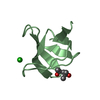
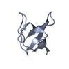
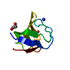
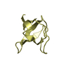
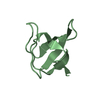
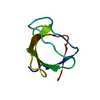
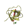
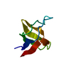
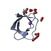
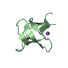
[English] 日本語
Yorodumi
- PDB-3ua6: Crystal Structure of the Human Fyn SH3 domain -

+
Open data


ID or keywords:

Loading...

-
Basic information

Entry
Database: PDB / ID: 3ua6
TitleCrystal Structure of the Human Fyn SH3 domain
ComponentsTyrosine-protein kinase Fyn
KeywordsTRANSFERASE / beta barrel
Function / homology
Function and homology information


negative regulation of hydrogen peroxide biosynthetic process / response to singlet oxygen / Reelin signalling pathway / perinuclear endoplasmic reticulum / NTRK2 activates RAC1 / growth factor receptor binding / regulation of glutamate receptor signaling pathway / heart process / Activated NTRK2 signals through FYN / SEMA3A-Plexin repulsion signaling by inhibiting Integrin adhesion ...negative regulation of hydrogen peroxide biosynthetic process / response to singlet oxygen / Reelin signalling pathway / perinuclear endoplasmic reticulum / NTRK2 activates RAC1 / growth factor receptor binding / regulation of glutamate receptor signaling pathway / heart process / Activated NTRK2 signals through FYN / SEMA3A-Plexin repulsion signaling by inhibiting Integrin adhesion / regulation of calcium ion import across plasma membrane / reelin-mediated signaling pathway / Platelet Adhesion to exposed collagen / CRMPs in Sema3A signaling / G protein-coupled glutamate receptor signaling pathway / FLT3 signaling through SRC family kinases / cellular response to L-glutamate / activated T cell proliferation / positive regulation of protein localization to membrane / type 5 metabotropic glutamate receptor binding / Nef and signal transduction / CD4 receptor binding / negative regulation of dendritic spine maintenance / feeding behavior / Co-stimulation by CD28 / Nephrin family interactions / DCC mediated attractive signaling / natural killer cell activation / EPH-Ephrin signaling / Ephrin signaling / CD28 dependent Vav1 pathway / dendritic spine maintenance / cellular response to peptide hormone stimulus / tau-protein kinase activity / Regulation of KIT signaling / leukocyte migration / phospholipase activator activity / Co-inhibition by CTLA4 / EPHA-mediated growth cone collapse / dendrite morphogenesis / peptide hormone receptor binding / CD8 receptor binding / Dectin-2 family / stimulatory C-type lectin receptor signaling pathway / Fc-gamma receptor signaling pathway involved in phagocytosis / PECAM1 interactions / response to amyloid-beta / cellular response to glycine / FCGR activation / Sema3A PAK dependent Axon repulsion / EPH-ephrin mediated repulsion of cells / glial cell projection / CD28 dependent PI3K/Akt signaling / positive regulation of protein targeting to membrane / Role of LAT2/NTAL/LAB on calcium mobilization / alpha-tubulin binding / negative regulation of extrinsic apoptotic signaling pathway in absence of ligand / vascular endothelial growth factor receptor signaling pathway / ephrin receptor signaling pathway / detection of mechanical stimulus involved in sensory perception of pain / postsynaptic density, intracellular component / cellular response to transforming growth factor beta stimulus / forebrain development / negative regulation of oxidative stress-induced intrinsic apoptotic signaling pathway / phospholipase binding / T cell receptor binding / phosphatidylinositol 3-kinase binding / GPVI-mediated activation cascade / T cell costimulation / cellular response to platelet-derived growth factor stimulus / ephrin receptor binding / Signaling by ERBB2 / EPHB-mediated forward signaling / negative regulation of protein ubiquitination / NCAM signaling for neurite out-growth / CD209 (DC-SIGN) signaling / FCGR3A-mediated IL10 synthesis / Regulation of signaling by CBL / axon guidance / Antigen activates B Cell Receptor (BCR) leading to generation of second messengers / cell surface receptor protein tyrosine kinase signaling pathway / Signaling by phosphorylated juxtamembrane, extracellular and kinase domain KIT mutants / negative regulation of angiogenesis / Cell surface interactions at the vascular wall / learning / actin filament / FCGR3A-mediated phagocytosis / non-membrane spanning protein tyrosine kinase activity / non-specific protein-tyrosine kinase / negative regulation of inflammatory response to antigenic stimulus / protein catabolic process / peptidyl-tyrosine phosphorylation / G protein-coupled receptor binding / Signaling by SCF-KIT / negative regulation of protein catabolic process / modulation of chemical synaptic transmission / positive regulation of neuron projection development / Signaling by CSF1 (M-CSF) in myeloid cells / tau protein binding / positive regulation of protein localization to nucleus
Similarity search - Function
: / Fyn/Yrk, SH3 domain / SH3 Domains / : / SH3 domain / SH2 domain / Src homology 2 (SH2) domain profile. / Src homology 2 domains / SH2 domain / Src homology 3 domains ...: / Fyn/Yrk, SH3 domain / SH3 Domains / : / SH3 domain / SH2 domain / Src homology 2 (SH2) domain profile. / Src homology 2 domains / SH2 domain / Src homology 3 domains / SH3 type barrels. / SH2 domain superfamily / SH3-like domain superfamily / Src homology 3 (SH3) domain profile. / SH3 domain / Tyrosine-protein kinase, catalytic domain / Tyrosine kinase, catalytic domain / Tyrosine protein kinases specific active-site signature. / Tyrosine-protein kinase, active site / Serine-threonine/tyrosine-protein kinase, catalytic domain / Protein tyrosine and serine/threonine kinase / Roll / Protein kinase, ATP binding site / Protein kinases ATP-binding region signature. / Protein kinase domain profile. / Protein kinase domain / Protein kinase-like domain superfamily / Mainly Beta
Similarity search - Domain/homology
FORMIC ACID / Tyrosine-protein kinase Fyn
Similarity search - Component
Biological speciesHomo sapiens (human)
MethodX-RAY DIFFRACTION / MOLECULAR REPLACEMENT / molecular replacement / Resolution: 1.85 Å
AuthorsCamara-Artigas, A. / Martin-Garcia, J.M.
CitationJournal: Acta Crystallogr.,Sect.D / Year: 2012
Title: The promiscuous binding of the Fyn SH3 domain to a peptide from the NS5A protein.
Authors: Martin-Garcia, J.M. / Luque, I. / Ruiz-Sanz, J. / Camara-Artigas, A.
History
DepositionOct 21, 2011Deposition site: RCSB / Processing site: RCSB
Revision 1.0Jul 25, 2012Provider: repository / Type: Initial release
Revision 1.1Jan 23, 2013Group: Database references
Revision 1.2Sep 13, 2023Group: Data collection / Database references ...Data collection / Database references / Derived calculations / Refinement description
Category: chem_comp_atom / chem_comp_bond ...chem_comp_atom / chem_comp_bond / database_2 / pdbx_initial_refinement_model / pdbx_struct_conn_angle / struct_conn / struct_ncs_dom_lim / struct_ref_seq_dif / struct_site
Item: _database_2.pdbx_DOI / _database_2.pdbx_database_accession ..._database_2.pdbx_DOI / _database_2.pdbx_database_accession / _pdbx_struct_conn_angle.ptnr1_auth_asym_id / _pdbx_struct_conn_angle.ptnr1_auth_comp_id / _pdbx_struct_conn_angle.ptnr1_auth_seq_id / _pdbx_struct_conn_angle.ptnr1_label_asym_id / _pdbx_struct_conn_angle.ptnr1_label_comp_id / _pdbx_struct_conn_angle.ptnr1_label_seq_id / _pdbx_struct_conn_angle.ptnr3_auth_asym_id / _pdbx_struct_conn_angle.ptnr3_auth_comp_id / _pdbx_struct_conn_angle.ptnr3_auth_seq_id / _pdbx_struct_conn_angle.ptnr3_label_asym_id / _pdbx_struct_conn_angle.ptnr3_label_comp_id / _pdbx_struct_conn_angle.ptnr3_label_seq_id / _pdbx_struct_conn_angle.value / _struct_conn.pdbx_dist_value / _struct_conn.ptnr1_auth_asym_id / _struct_conn.ptnr1_auth_comp_id / _struct_conn.ptnr1_auth_seq_id / _struct_conn.ptnr1_label_asym_id / _struct_conn.ptnr1_label_atom_id / _struct_conn.ptnr1_label_comp_id / _struct_conn.ptnr1_label_seq_id / _struct_conn.ptnr2_auth_asym_id / _struct_conn.ptnr2_auth_comp_id / _struct_conn.ptnr2_auth_seq_id / _struct_conn.ptnr2_label_asym_id / _struct_conn.ptnr2_label_atom_id / _struct_conn.ptnr2_label_comp_id / _struct_conn.ptnr2_label_seq_id / _struct_ncs_dom_lim.beg_auth_comp_id / _struct_ncs_dom_lim.beg_label_asym_id / _struct_ncs_dom_lim.beg_label_comp_id / _struct_ncs_dom_lim.beg_label_seq_id / _struct_ncs_dom_lim.end_auth_comp_id / _struct_ncs_dom_lim.end_label_asym_id / _struct_ncs_dom_lim.end_label_comp_id / _struct_ncs_dom_lim.end_label_seq_id / _struct_ref_seq_dif.details / _struct_site.pdbx_auth_asym_id / _struct_site.pdbx_auth_comp_id / _struct_site.pdbx_auth_seq_id

-
Structure visualization

Structure viewerMolecule:
MolmilJmol/JSmol

Downloads & links

-
Assembly

Deposited unit
A: Tyrosine-protein kinase Fyn
B: Tyrosine-protein kinase Fyn
hetero molecules


Theoretical massNumber of molelcules
Total (without water)14,87313
Polymers14,3442
Non-polymers52911
Water1,35175
1
A: Tyrosine-protein kinase Fyn
hetero molecules


Theoretical massNumber of molelcules
Total (without water)7,63210
Polymers7,1721
Non-polymers4609
Water181
TypeNameSymmetry operationNumber
identity operation1_555x,y,z1
2
B: Tyrosine-protein kinase Fyn
hetero molecules


Theoretical massNumber of molelcules
Total (without water)7,2413
Polymers7,1721
Non-polymers692
Water181
TypeNameSymmetry operationNumber
identity operation1_555x,y,z1
Unit cell
Length a, b, c (Å)72.293, 47.074, 42.467
Angle α, β, γ (deg.)90.000, 98.520, 90.000
Int Tables number5
Space group name H-MC121
Noncrystallographic symmetry (NCS)NCS domain:
IDEns-IDDetails
11A
21B

NCS domain segments:

Ens-ID: 1

Dom-IDComponent-IDBeg auth comp-IDBeg label comp-IDEnd auth comp-IDEnd label comp-IDRefine codeAuth asym-IDLabel asym-IDAuth seq-IDLabel seq-ID
11THRTHRASNASN2AA85 - 1136 - 34
21THRTHRASNASN2BB85 - 1136 - 34
12ASPASPPROPRO4AA118 - 14039 - 61
22ASPASPPROPRO4BB118 - 14039 - 61

-
Components

#1: Protein Tyrosine-protein kinase Fyn / Proto-oncogene Syn / Proto-oncogene c-Fyn / Src-like kinase / SLK / p59-Fyn


Mass: 7171.764 Da / Num. of mol.: 2 / Fragment: SH3 domain (UNP residues 81-143)
Source method: isolated from a genetically manipulated source
Source: (gene. exp.) Homo sapiens (human) / Gene: FYN / Plasmid: pET3d / Production host: Escherichia coli (E. coli) / Strain (production host): BL21(DE3)
References: UniProt: P06241, non-specific protein-tyrosine kinase
#2: Chemical ChemComp-GOL / GLYCEROL / GLYCERIN / PROPANE-1,2,3-TRIOL


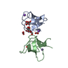
Mass: 92.094 Da / Num. of mol.: 2 / Source method: obtained synthetically / Formula: C3H8O3
#3: Chemical
ChemComp-FMT / FORMIC ACID


Mass: 46.025 Da / Num. of mol.: 6 / Source method: obtained synthetically / Formula: CH2O2
#4: Chemical ChemComp-NA / SODIUM ION


Mass: 22.990 Da / Num. of mol.: 3 / Source method: obtained synthetically / Formula: Na
#5: Water ChemComp-HOH / water


Mass: 18.015 Da / Num. of mol.: 75 / Source method: isolated from a natural source / Formula: H2O

-
Experimental details

-
Experiment

ExperimentMethod: X-RAY DIFFRACTION / Number of used crystals: 1

-
Sample preparation

CrystalDensity Matthews: 2.49 Å3/Da / Density % sol: 50.62 %
Crystal growTemperature: 298 K / Method: vapor diffusion, hanging drop / pH: 6
Details: 5.5 M sodium formate, 0.1 M MES, pH 6.0, VAPOR DIFFUSION, HANGING DROP, temperature 298K

-
Data collection

DiffractionMean temperature: 100 K
Diffraction sourceSource: ROTATING ANODE / Type: BRUKER AXS MICROSTAR / Wavelength: 1.54 Å
DetectorType: Bruker Microfocus (Montel Optics) / Detector: CCD / Date: Nov 1, 2006
RadiationProtocol: SINGLE WAVELENGTH / Monochromatic (M) / Laue (L): M / Scattering type: x-ray
Radiation wavelengthWavelength: 1.54 Å / Relative weight: 1
ReflectionResolution: 1.85→41.998 Å / Num. all: 12182 / Num. obs: 11459 / % possible obs: 94.1 % / Observed criterion σ(F): 0 / Observed criterion σ(I): 0 / Redundancy: 2.49 % / Biso Wilson estimate: 21.92 Å2 / Rmerge(I) obs: 0.0274 / Rsym value: 0.0274 / Net I/σ(I): 21.66
Reflection shellResolution: 1.85→1.95 Å / Redundancy: 0.87 % / Rmerge(I) obs: 0.1511 / Mean I/σ(I) obs: 3.78 / Num. unique all: 1109 / Rsym value: 0.1511 / % possible all: 72.4

-
Phasing

PhasingMethod: molecular replacement
Phasing MR
Highest resolutionLowest resolution
Rotation2.02 Å42 Å
Translation2.02 Å42 Å

-
Processing

Software
NameVersionClassificationNB
SAINTdata scaling
SCALAdata scaling
MOLREPphasing
REFMAC5.5.0109refinement
PDB_EXTRACT3.1data extraction
APEXdata collection
SAINTdata reduction
RefinementMethod to determine structure: MOLECULAR REPLACEMENT
Starting model: PDB ENTRY 1SHF
Resolution: 1.85→19.66 Å / Cor.coef. Fo:Fc: 0.958 / Cor.coef. Fo:Fc free: 0.921 / Occupancy max: 1 / Occupancy min: 0.5 / SU B: 6.052 / SU ML: 0.086 / Cross valid method: THROUGHOUT / σ(F): 0 / σ(I): 0 / ESU R: 0.137 / ESU R Free: 0.148 / Stereochemistry target values: MAXIMUM LIKELIHOOD
Details: HYDROGENS HAVE BEEN ADDED IN THE RIDING POSITIONS U VALUES : RESIDUAL ONLY
RfactorNum. reflection% reflectionSelection details
Rfree0.2464 544 4.8 %RANDOM
Rwork0.177 ---
all0.1801 12170 --
obs0.1801 11447 94.06 %-
Solvent computationIon probe radii: 0.8 Å / Shrinkage radii: 0.8 Å / VDW probe radii: 1.4 Å / Solvent model: MASK
Displacement parametersBiso max: 67.82 Å2 / Biso mean: 25.454 Å2 / Biso min: 2 Å2
Baniso -1Baniso -2Baniso -3
1--0.05 Å20 Å2-0.06 Å2
2--0.53 Å20 Å2
3----0.5 Å2
Refinement stepCycle: LAST / Resolution: 1.85→19.66 Å
ProteinNucleic acidLigandSolventTotal
Num. atoms943 0 33 75 1051
Refine LS restraints
Refine-IDTypeDev idealDev ideal targetNumber
X-RAY DIFFRACTIONr_bond_refined_d0.020.0221018
X-RAY DIFFRACTIONr_angle_refined_deg1.9781.9471377
X-RAY DIFFRACTIONr_dihedral_angle_1_deg7.3045121
X-RAY DIFFRACTIONr_dihedral_angle_2_deg39.36524.44454
X-RAY DIFFRACTIONr_dihedral_angle_3_deg13.3815151
X-RAY DIFFRACTIONr_dihedral_angle_4_deg11.186155
X-RAY DIFFRACTIONr_chiral_restr0.1390.2142
X-RAY DIFFRACTIONr_gen_planes_refined0.0080.021795
X-RAY DIFFRACTIONr_mcbond_it2.0061.5603
X-RAY DIFFRACTIONr_mcangle_it2.9332953
X-RAY DIFFRACTIONr_scbond_it4.3543415
X-RAY DIFFRACTIONr_scangle_it6.7314.5422
Refine LS restraints NCS

Dom-ID: 1 / Auth asym-ID: A / Ens-ID: 1 / Refine-ID: X-RAY DIFFRACTION

NumberTypeRms dev position (Å)Weight position
114TIGHT POSITIONAL0.50.05
293MEDIUM POSITIONAL0.760.5
114TIGHT THERMAL1.790.5
293MEDIUM THERMAL2.192
LS refinement shellResolution: 1.85→1.898 Å / Total num. of bins used: 20
RfactorNum. reflection% reflection
Rfree0.247 25 -
Rwork0.188 454 -
all-479 -
obs--54.06 %
Refinement TLS params.

Method: refined / Refine-ID: X-RAY DIFFRACTION

IDL112)L122)L132)L222)L232)L332)S11 (Å °)S12 (Å °)S13 (Å °)S21 (Å °)S22 (Å °)S23 (Å °)S31 (Å °)S32 (Å °)S33 (Å °)T112)T122)T132)T222)T232)T332)Origin x (Å)Origin y (Å)Origin z (Å)
12.8237-0.6960.21981.5698-0.4343.7608-0.00250.08570.04760.01510.0271-0.02150.01780.1312-0.02470.01480.012-0.00270.0245-0.01560.0284-7.656-0.41-10.494
22.2479-1.4578-0.18252.09040.57763.3265-0.0664-0.00520.0465-0.0101-0.05520.1065-0.20140.02140.12170.04510.0169-0.00760.02070.00470.0285-22.83215.502-9.795
Refinement TLS group
IDRefine-IDRefine TLS-IDAuth asym-IDAuth seq-ID
1X-RAY DIFFRACTION1A85 - 141
2X-RAY DIFFRACTION2B83 - 142

+
About Yorodumi

-
News

-
Feb 9, 2022. New format data for meta-information of EMDB entries

New format data for meta-information of EMDB entries

  • Version 3 of the EMDB header file is now the official format.
  • The previous official version 1.9 will be removed from the archive.

Related info.:EMDB header

External links:wwPDB to switch to version 3 of the EMDB data model

-
Aug 12, 2020. Covid-19 info

Covid-19 info

URL: https://pdbj.org/emnavi/covid19.php

New page: Covid-19 featured information page in EM Navigator.

Related info.:Covid-19 info / Mar 5, 2020. Novel coronavirus structure data

+
Mar 5, 2020. Novel coronavirus structure data

Novel coronavirus structure data

Related info.:Yorodumi Speices / Aug 12, 2020. Covid-19 info

External links:COVID-19 featured content - PDBj / Molecule of the Month (242):Coronavirus Proteases

+
Jan 31, 2019. EMDB accession codes are about to change! (news from PDBe EMDB page)

EMDB accession codes are about to change! (news from PDBe EMDB page)

  • The allocation of 4 digits for EMDB accession codes will soon come to an end. Whilst these codes will remain in use, new EMDB accession codes will include an additional digit and will expand incrementally as the available range of codes is exhausted. The current 4-digit format prefixed with “EMD-” (i.e. EMD-XXXX) will advance to a 5-digit format (i.e. EMD-XXXXX), and so on. It is currently estimated that the 4-digit codes will be depleted around Spring 2019, at which point the 5-digit format will come into force.
  • The EM Navigator/Yorodumi systems omit the EMD- prefix.

Related info.:Q: What is EMD? / ID/Accession-code notation in Yorodumi/EM Navigator

External links:EMDB Accession Codes are Changing Soon! / Contact to PDBj

+
Jul 12, 2017. Major update of PDB

Major update of PDB

  • wwPDB released updated PDB data conforming to the new PDBx/mmCIF dictionary.
  • This is a major update changing the version number from 4 to 5, and with Remediation, in which all the entries are updated.
  • In this update, many items about electron microscopy experimental information are reorganized (e.g. em_software).
  • Now, EM Navigator and Yorodumi are based on the updated data.

External links:wwPDB Remediation / Enriched Model Files Conforming to OneDep Data Standards Now Available in the PDB FTP Archive

-
Yorodumi

Thousand views of thousand structures

  • Yorodumi is a browser for structure data from EMDB, PDB, SASBDB, etc.
  • This page is also the successor to EM Navigator detail page, and also detail information page/front-end page for Omokage search.
  • The word "yorodu" (or yorozu) is an old Japanese word meaning "ten thousand". "mi" (miru) is to see.

Related info.:EMDB / PDB / SASBDB / Comparison of 3 databanks / Yorodumi Search / Aug 31, 2016. New EM Navigator & Yorodumi / Yorodumi Papers / Jmol/JSmol / Function and homology information / Changes in new EM Navigator and Yorodumi

Read more