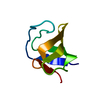
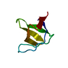
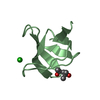
Entry Database : PDB / ID : 5fwcTitle Human Spectrin SH3 domain D48G, E7A, K60A SPECTRIN ALPHA CHAIN, NON-ERYTHROCYTIC 1 Keywords / Function / homology Function Domain/homology Component
/ / / / / / / / / / / / / / / / / / / / / / / / / / / / / / / / / / / / / / / / / / / / / / / / / / / / / / / / / / / / / / / / / / / / / / / / Biological species HOMO SAPIENS (human)Method / / / Resolution : 1.4 Å Authors Gallego, P. / Navarro, S. / Ventura, S. / Reverter, D. Journal : To be Published Title : Human Spectrin SH3 Domain D48G, E7A, K60AAuthors : Navarro, S. / Gallego, P. / Diaz, M. / Ventura, S. / Reverter, D. History Deposition Feb 12, 2016 Deposition site / Processing site Revision 1.0 Dec 28, 2016 Provider / Type Revision 1.1 Jan 10, 2024 Group Data collection / Database references ... Data collection / Database references / Derived calculations / Other / Refinement description Category chem_comp_atom / chem_comp_bond ... chem_comp_atom / chem_comp_bond / database_2 / pdbx_database_status / pdbx_initial_refinement_model / struct_site Item _database_2.pdbx_DOI / _database_2.pdbx_database_accession ... _database_2.pdbx_DOI / _database_2.pdbx_database_accession / _pdbx_database_status.status_code_sf / _struct_site.pdbx_auth_asym_id / _struct_site.pdbx_auth_comp_id / _struct_site.pdbx_auth_seq_id
Show all Show less
 Movie
Movie Controller


 Open data
 Basic information
 Components
 Keywords
 Function and homology information
 HOMO SAPIENS (human)
 X-RAY DIFFRACTION /
  Authors
 Citation
 Structure visualization
 Downloads & links
 Other downloads


 PDBj
PDBj


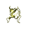
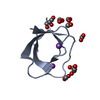
 Assembly
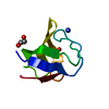


 Mass: 62.068 Da / Num. of mol.: 2 / Source method: obtained synthetically / Formula: C2H6O2
 Mass: 18.038 Da / Num. of mol.: 1 / Source method: obtained synthetically / Formula: H4N
 Mass: 18.015 Da / Num. of mol.: 55 / Source method: isolated from a natural source / Formula: H2O
 Sample preparation
 / Beamline: XALOC / Wavelength: 0.979493
  Processing