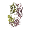
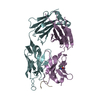
Mass: 25965.783 Da / Num. of mol.: 1 Source method: isolated from a genetically manipulated source Source: (gene. exp.) Homo sapiens (human) / Gene: IGHG1 / Cell line (production host): HEK293F / Production host: Homo sapiens (human) / References: UniProt: P01857
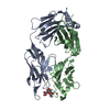
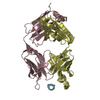
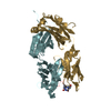
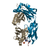
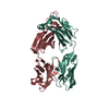
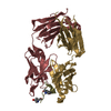
#2: Antibody
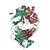
APE1551AbFablightchain
Mass: 24342.928 Da / Num. of mol.: 1 Source method: isolated from a genetically manipulated source Source: (gene. exp.) Homo sapiens (human) / Cell line (production host): HEK293F / Production host: Homo sapiens (human) / References: UniProt: Q8TCD0
Resolution: 1.6→1.63 Å / Redundancy: 5 % / Rmerge(I) obs: 0.623 / Mean I/σ(I) obs: 2.4 / Num. unique all: 3210 / % possible all: 96.3
-
Processing
Software
Name
Version
Classification
Web-Ice
datacollection
PHASER
phasing
REFMAC
5.7.0029
refinement
HKL-2000
datareduction
HKL-2000
datascaling
Refinement
Method to determine structure: MOLECULAR REPLACEMENT Starting model: APE1531 Fab Resolution: 1.602→45.58 Å / Cor.coef. Fo:Fc: 0.973 / Cor.coef. Fo:Fc free: 0.962 / SU B: 3.701 / SU ML: 0.064 / Cross valid method: THROUGHOUT / ESU R: 0.083 / ESU R Free: 0.085 / Stereochemistry target values: MAXIMUM LIKELIHOOD / Details: HYDROGENS HAVE BEEN ADDED IN THE RIDING POSITIONS
Rfactor
Num. reflection
% reflection
Selection details
Rfree
0.19767
3280
5.1 %
RANDOM
Rwork
0.16628
-
-
-
obs
0.16787
61414
97.83 %
-
Solvent computation
Ion probe radii: 0.8 Å / Shrinkage radii: 0.8 Å / VDW probe radii: 1.2 Å / Solvent model: MASK
 Movie
Movie Controller

 Yorodumi
 Open data
 Basic information
 Components
 Keywords
 Function and homology information
 Homo sapiens (human)
 X-RAY DIFFRACTION /
  Authors
 Citation
 Structure visualization
 Downloads & links
 Other downloads
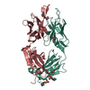


 PDBj
PDBj


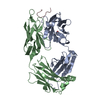
 Assembly
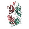
 Mass: 18.015 Da / Num. of mol.: 434 / Source method: isolated from a natural source / Formula: H2O
 Sample preparation
 / Beamline: BL12-2 / Wavelength: 0.97946 Å
 Processing