 Movie
Movie Controller

[English] 日本語
 Yorodumi
- PDB-2or9: The structure of the anti-c-myc antibody 9E10 Fab fragment/epitop... -
+ Open data

- Basic information
| Entry | Database: PDB / ID: 2or9 | ||||||
|---|---|---|---|---|---|---|---|
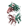
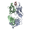
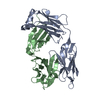
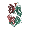
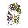
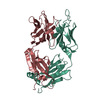
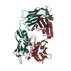
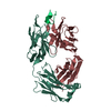
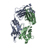
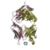
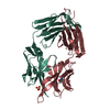
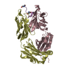
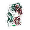
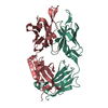
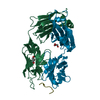
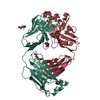
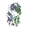
| Title | The structure of the anti-c-myc antibody 9E10 Fab fragment/epitope peptide complex reveals a novel binding mode dominated by the heavy chain hypervariable loops | ||||||
 Components Components |
| ||||||
 Keywords Keywords | IMMUNE SYSTEM / ANTIGEN-ANTIBODY COMPLEX / ANTIGEN RECOGNITION / myc-tag / long CDR H3 | ||||||
| Function / homology |  Function and homology information Function and homology informationpositive regulation of metanephric cap mesenchymal cell proliferation / positive regulation of acinar cell proliferation / acinar cell proliferation / SCF ubiquitin ligase complex binding / NK T cell proliferation / Myc-Max complex / regulation of somatic stem cell population maintenance / regulation of cell cycle process / RNA polymerase II transcription repressor complex / Binding of TCF/LEF:CTNNB1 to target gene promoters ...positive regulation of metanephric cap mesenchymal cell proliferation / positive regulation of acinar cell proliferation / acinar cell proliferation / SCF ubiquitin ligase complex binding / NK T cell proliferation / Myc-Max complex / regulation of somatic stem cell population maintenance / regulation of cell cycle process / RNA polymerase II transcription repressor complex / Binding of TCF/LEF:CTNNB1 to target gene promoters / cellular response to interferon-alpha / positive regulation of B cell apoptotic process / RUNX3 regulates WNT signaling / TFAP2 (AP-2) family regulates transcription of cell cycle factors / myotube differentiation / negative regulation of transcription initiation by RNA polymerase II / negative regulation of cell division / Regulation of CDH1 mRNA translation by microRNAs / negative regulation of monocyte differentiation / detection of mechanical stimulus involved in sensory perception of sound / response to growth factor / B cell apoptotic process / response to alkaloid / transcription regulator activator activity / Transcription of E2F targets under negative control by DREAM complex / fibroblast apoptotic process / negative regulation of stress-activated MAPK cascade / Regulation of NFE2L2 gene expression / protein-DNA complex disassembly / positive regulation of mesenchymal cell proliferation / skeletal system morphogenesis / regulation of telomere maintenance / Signaling by ALK / branching involved in ureteric bud morphogenesis / middle ear morphogenesis / rRNA metabolic process / negative regulation of gene expression via chromosomal CpG island methylation / E-box binding / pigmentation / positive regulation of telomere maintenance / skeletal muscle cell differentiation / chromosome organization / positive regulation of transcription initiation by RNA polymerase II / positive regulation of intrinsic apoptotic signaling pathway by p53 class mediator / Cyclin E associated events during G1/S transition / core promoter sequence-specific DNA binding / Cyclin A:Cdk2-associated events at S phase entry / negative regulation of fibroblast proliferation / ERK1 and ERK2 cascade / positive regulation of epithelial cell proliferation / transcription coregulator binding / G1/S transition of mitotic cell cycle / euchromatin / SMAD2/SMAD3:SMAD4 heterotrimer regulates transcription / protein processing / positive regulation of miRNA transcription / MAPK6/MAPK4 signaling / DNA-binding transcription repressor activity, RNA polymerase II-specific / NOTCH1 Intracellular Domain Regulates Transcription / cellular response to xenobiotic stimulus / spindle / Constitutive Signaling by NOTCH1 PEST Domain Mutants / Constitutive Signaling by NOTCH1 HD+PEST Domain Mutants / positive regulation of fibroblast proliferation / intrinsic apoptotic signaling pathway in response to DNA damage / Transcriptional regulation of granulopoiesis / Wnt signaling pathway / cellular response to UV / MAPK cascade / regulation of gene expression / Interleukin-4 and Interleukin-13 signaling / DNA-binding transcription activator activity, RNA polymerase II-specific / transcription by RNA polymerase II / cellular response to hypoxia / Estrogen-dependent gene expression / DNA-binding transcription factor binding / intracellular iron ion homeostasis / DNA-binding transcription factor activity, RNA polymerase II-specific / protein dimerization activity / Ub-specific processing proteases / nuclear body / RNA polymerase II cis-regulatory region sequence-specific DNA binding / chromatin remodeling / response to xenobiotic stimulus / axon / positive regulation of cell population proliferation / DNA damage response / ubiquitin protein ligase binding / positive regulation of gene expression / regulation of transcription by RNA polymerase II / negative regulation of apoptotic process / positive regulation of DNA-templated transcription / chromatin / protein-containing complex binding / nucleolus / perinuclear region of cytoplasm / negative regulation of transcription by RNA polymerase II / positive regulation of transcription by RNA polymerase II / protein-containing complex / DNA binding Similarity search - Function | ||||||
| Biological species |  | ||||||
| Method |  X-RAY DIFFRACTION / SYNCHROTRON / MOLECULAR REPLACEMENT / Resolution: 2.7 Å X-RAY DIFFRACTION / SYNCHROTRON / MOLECULAR REPLACEMENT / Resolution: 2.7 Å | ||||||
 Authors Authors | Krauss, N. / Scheerer, P. / Hoehne, W. | ||||||
 Citation Citation | Journal: Proteins / Year: 2008 Title: The structure of the anti-c-myc antibody 9E10 Fab fragment/epitope peptide complex reveals a novel binding mode dominated by the heavy chain hypervariable loops. Authors: Krauss, N. / Wessner, H. / Welfle, K. / Welfle, H. / Scholz, C. / Seifert, M. / Zubow, K. / Ay, J. / Hahn, M. / Scheerer, P. / Skerra, A. / Hohne, W. | ||||||
| History |
|
- Structure visualization
| Structure viewer | Molecule: MolmilJmol/JSmol |
|---|
- Downloads & links
-Download
| PDBx/mmCIF format | 2or9.cif.gz | 186.7 KB | Display | PDBx/mmCIF format |
|---|---|---|---|---|
| PDB format | pdb2or9.ent.gz | 149 KB | Display | PDB format |
| PDBx/mmJSON format | 2or9.json.gz | Tree view | PDBx/mmJSON format | |
| Others |  Other downloads Other downloads |
-Validation report
| Arichive directory | https://data.pdbj.org/pub/pdb/validation_reports/or/2or9ftp://data.pdbj.org/pub/pdb/validation_reports/or/2or9 | HTTPS FTP |
|---|
-Related structure data
| Related structure data | 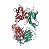 2orbC  1cloS C: citing same article ( S: Starting model for refinement |
|---|---|
| Similar structure data |


-Links
 PDBj
PDBj


- Assembly
| Deposited unit | 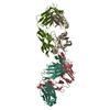
| ||||||||
|---|---|---|---|---|---|---|---|---|---|
| 1 | 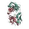
| ||||||||
| 2 | 
| ||||||||
| Unit cell |
|
-Components
| #1: Antibody | Mass: 24001.543 Da / Num. of mol.: 2 / Fragment: light chain of antigen binding fragment, Fab / Source method: isolated from a natural source / Source: (natural) #2: Antibody | Mass: 24899.799 Da / Num. of mol.: 2 / Fragment: heavy chain of antigen binding fragment, Fab / Source method: isolated from a natural source / Source: (natural) #3: Protein/peptide | | Mass: 1318.407 Da / Num. of mol.: 1 / Source method: obtained synthetically Details: synthetic peptide derived from the human c-myc proto-oncoprotein References: UniProt: P01106*PLUS #4: Water | ChemComp-HOH / |  Mass: 18.015 Da / Num. of mol.: 282 / Source method: isolated from a natural source / Formula: H2O Mass: 18.015 Da / Num. of mol.: 282 / Source method: isolated from a natural source / Formula: H2OHas protein modification | Y | |
|---|
-Experimental details
-Experiment
| Experiment | Method: X-RAY DIFFRACTION / Number of used crystals: 1 |
|---|
- Sample preparation
| Crystal | Density Matthews: 2.88 Å3/Da / Density % sol: 57.25 % |
|---|---|
| Crystal grow | Temperature: 291 K / Method: vapor diffusion, hanging drop / pH: 5 Details: 12% PEG 4000, 0.1 to 0.2 M sodium acetate, 0.02% sodium azide, pH 5.0, VAPOR DIFFUSION, HANGING DROP, temperature 291K |
-Data collection
| Diffraction | Mean temperature: 100 K |
|---|---|
| Diffraction source | Source: SYNCHROTRON / Site: EMBL/DESY, HAMBURG  / Beamline: X11 / Wavelength: 0.91 / Beamline: X11 / Wavelength: 0.91 |
| Detector | Type: MAR CCD 165 mm / Detector: CCD / Date: Aug 25, 2000 |
| Radiation | Monochromator: TRIANGULAR MONOCHROMATOR / Protocol: SINGLE WAVELENGTH / Monochromatic (M) / Laue (L): M / Scattering type: x-ray |
| Radiation wavelength | Wavelength: 0.91 Å / Relative weight: 1 |
| Reflection | Resolution: 2.7→20 Å / Num. all: 31926 / Num. obs: 31926 / % possible obs: 99.5 % / Observed criterion σ(F): 0 / Observed criterion σ(I): -3 / Redundancy: 3.6 % / Biso Wilson estimate: 73.2 Å2 / Rsym value: 0.04 / Net I/σ(I): 26.7 |
| Reflection shell | Resolution: 2.7→2.8 Å / Redundancy: 3.1 % / Mean I/σ(I) obs: 4.1 / Num. unique all: 3088 / Rsym value: 0.25 / % possible all: 97.4 |
- Processing
| Software |
| |||||||||||||||||||||||||
|---|---|---|---|---|---|---|---|---|---|---|---|---|---|---|---|---|---|---|---|---|---|---|---|---|---|---|
| Refinement | Method to determine structure: MOLECULAR REPLACEMENT Starting model: PDB entry 1CLO Resolution: 2.7→20 Å / Isotropic thermal model: isotropic / Cross valid method: THROUGHOUT / σ(F): 0 / Stereochemistry target values: Engh & Huber
| |||||||||||||||||||||||||
| Displacement parameters | Biso mean: 59.1 Å2 | |||||||||||||||||||||||||
| Refine analyze |
| |||||||||||||||||||||||||
| Refinement step | Cycle: LAST / Resolution: 2.7→20 Å
| |||||||||||||||||||||||||
| Refine LS restraints |
| |||||||||||||||||||||||||
| LS refinement shell | Resolution: 2.7→2.8 Å
|
