 Movie
Movie Controller

[English] 日本語
 Yorodumi
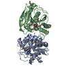
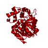
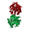
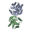
- PDB-4lxg: Crystal structure of DxnB2, a carbon - carbon bond hydrolase from... -
+ Open data

- Basic information
| Entry | Database: PDB / ID: 4lxg | ||||||
|---|---|---|---|---|---|---|---|
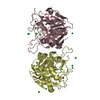
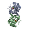
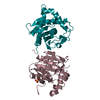
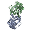
| Title | Crystal structure of DxnB2, a carbon - carbon bond hydrolase from Sphingomonas wittichii RW1 | ||||||
 Components Components | MCP Hydrolase | ||||||
 Keywords Keywords | HYDROLASE / Carbon-carbon bond hydrolase / Rossmann Fold / alpha/beta hydrolase fold / Cytosolic | ||||||
| Function / homology | Alpha/beta hydrolase family / Alpha/beta hydrolase fold-1 / Alpha/Beta hydrolase fold, catalytic domain / Alpha/Beta hydrolase fold / hydrolase activity / Rossmann fold / 3-Layer(aba) Sandwich / Alpha Beta / Alpha/beta hydrolase fold Function and homology information Function and homology information | ||||||
| Biological species |  Sphingomonas wittichii RW1 (bacteria) Sphingomonas wittichii RW1 (bacteria) | ||||||
| Method |  X-RAY DIFFRACTION / SYNCHROTRON / MOLECULAR REPLACEMENT / molecular replacement / Resolution: 2.22 Å X-RAY DIFFRACTION / SYNCHROTRON / MOLECULAR REPLACEMENT / molecular replacement / Resolution: 2.22 Å | ||||||
 Authors Authors | Bhowmik, S. / Bolin, J.T. | ||||||
 Citation Citation | Journal: Biochemistry / Year: 2013 Title: The Lid Domain of the MCP Hydrolase DxnB2 Contributes to the Reactivity toward Recalcitrant PCB Metabolites. Authors: Ruzzini, A.C. / Bhowmik, S. / Yam, K.C. / Ghosh, S. / Bolin, J.T. / Eltis, L.D. | ||||||
| History |
|
- Structure visualization
| Structure viewer | Molecule: MolmilJmol/JSmol |
|---|
- Downloads & links
-Download
| PDBx/mmCIF format | 4lxg.cif.gz | 119.6 KB | Display | PDBx/mmCIF format |
|---|---|---|---|---|
| PDB format | pdb4lxg.ent.gz | 93.4 KB | Display | PDB format |
| PDBx/mmJSON format | 4lxg.json.gz | Tree view | PDBx/mmJSON format | |
| Others |  Other downloads Other downloads |
-Validation report
| Arichive directory | https://data.pdbj.org/pub/pdb/validation_reports/lx/4lxgftp://data.pdbj.org/pub/pdb/validation_reports/lx/4lxg | HTTPS FTP |
|---|
-Related structure data
| Related structure data |  4lxhC 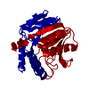 4lydC 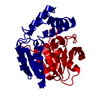 1j1iS C: citing same article ( S: Starting model for refinement |
|---|---|
| Similar structure data |

-Links
 PDBj
PDBj

- Assembly
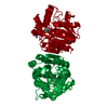
| Deposited unit | 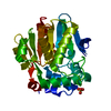
| ||||||||
|---|---|---|---|---|---|---|---|---|---|
| 1 | 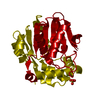
| ||||||||
| Unit cell |
|
-Components
| #1: Protein | Mass: 30224.531 Da / Num. of mol.: 1 Source method: isolated from a genetically manipulated source Source: (gene. exp.) Sphingomonas wittichii RW1 (bacteria) / Strain: RW1 / Gene: dxnB2, Swit_3055 / Production host: References: UniProt: A5VAT9, 2,6-dioxo-6-phenylhexa-3-enoate hydrolase | ||
|---|---|---|---|
| #2: Chemical |   Mass: 96.063 Da / Num. of mol.: 3 / Source method: obtained synthetically / Formula: SO4 Mass: 96.063 Da / Num. of mol.: 3 / Source method: obtained synthetically / Formula: SO4#3: Water | ChemComp-HOH / |  Mass: 18.015 Da / Num. of mol.: 81 / Source method: isolated from a natural source / Formula: H2O Mass: 18.015 Da / Num. of mol.: 81 / Source method: isolated from a natural source / Formula: H2O |
-Experimental details
-Experiment
| Experiment | Method: X-RAY DIFFRACTION / Number of used crystals: 1 |
|---|
- Sample preparation
| Crystal | Density Matthews: 3.47 Å3/Da / Density % sol: 64.58 % |
|---|---|
| Crystal grow | Temperature: 293 K / Method: vapor diffusion / pH: 8.2 Details: grid of ammonium sulfate (1.0-2.5 M) containing 0.1 M Tris, pH 8.2 and 6-10% ethanol, VAPOR DIFFUSION, temperature 293K |
-Data collection
| Diffraction | Mean temperature: 100 K |
|---|---|
| Diffraction source | Source: SYNCHROTRON / Site: APS  / Beamline: 14-BM-C / Wavelength: 1 Å / Beamline: 14-BM-C / Wavelength: 1 Å |
| Radiation | Protocol: SINGLE WAVELENGTH / Monochromatic (M) / Laue (L): M / Scattering type: x-ray |
| Radiation wavelength | Wavelength: 1 Å / Relative weight: 1 |
| Reflection | Resolution: 2.2→58.03 Å / Num. all: 22668 / Num. obs: 22668 / % possible obs: 99.5 % / Observed criterion σ(F): 2 / Observed criterion σ(I): 2 |
| Reflection shell | Resolution: 2.2→2.2 Å / % possible all: 99.5 |
-Phasing
| Phasing | Method: molecular replacement |
|---|
- Processing
| Software |
| |||||||||||||||||||||||||||||||||||||||||||||||||||||||||||||||||||||||||||
|---|---|---|---|---|---|---|---|---|---|---|---|---|---|---|---|---|---|---|---|---|---|---|---|---|---|---|---|---|---|---|---|---|---|---|---|---|---|---|---|---|---|---|---|---|---|---|---|---|---|---|---|---|---|---|---|---|---|---|---|---|---|---|---|---|---|---|---|---|---|---|---|---|---|---|---|---|
| Refinement | Method to determine structure: MOLECULAR REPLACEMENT Starting model: PDB ENTRY 1J1I Resolution: 2.22→36.46 Å / Cor.coef. Fo:Fc: 0.952 / Cor.coef. Fo:Fc free: 0.93 / Occupancy max: 1 / Occupancy min: 0.5 / SU B: 11.378 / SU ML: 0.139 / Cross valid method: THROUGHOUT / σ(F): 0 / ESU R: 0.19 / ESU R Free: 0.169 / Stereochemistry target values: MAXIMUM LIKELIHOOD Details: HYDROGENS HAVE BEEN USED IF PRESENT IN THE INPUT U VALUES: WITH TLS ADDED
| |||||||||||||||||||||||||||||||||||||||||||||||||||||||||||||||||||||||||||
| Solvent computation | Ion probe radii: 0.8 Å / Shrinkage radii: 0.8 Å / VDW probe radii: 1.2 Å / Solvent model: BABINET MODEL WITH MASK | |||||||||||||||||||||||||||||||||||||||||||||||||||||||||||||||||||||||||||
| Displacement parameters | Biso max: 119.64 Å2 / Biso mean: 52.9638 Å2 / Biso min: 31.43 Å2
| |||||||||||||||||||||||||||||||||||||||||||||||||||||||||||||||||||||||||||
| Refinement step | Cycle: LAST / Resolution: 2.22→36.46 Å
| |||||||||||||||||||||||||||||||||||||||||||||||||||||||||||||||||||||||||||
| Refine LS restraints |
| |||||||||||||||||||||||||||||||||||||||||||||||||||||||||||||||||||||||||||
| LS refinement shell | Resolution: 2.223→2.281 Å / Total num. of bins used: 20
| |||||||||||||||||||||||||||||||||||||||||||||||||||||||||||||||||||||||||||
| Refinement TLS params. | Method: refined / Refine-ID: X-RAY DIFFRACTION
| |||||||||||||||||||||||||||||||||||||||||||||||||||||||||||||||||||||||||||
| Refinement TLS group |
|
