 Movie
Movie Controller

[English] 日本語
 Yorodumi
- PDB-1j1i: Crystal structure of a His-tagged Serine Hydrolase Involved in th... -
+ Open data

- Basic information
| Entry | Database: PDB / ID: 1j1i | ||||||
|---|---|---|---|---|---|---|---|
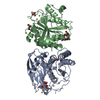
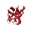
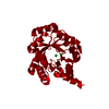
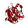
| Title | Crystal structure of a His-tagged Serine Hydrolase Involved in the Carbazole Degradation (CarC enzyme) | ||||||
 Components Components | meta cleavage compound hydrolase | ||||||
 Keywords Keywords | HYDROLASE / Carbazole degradation / meta cleavage product hydrolase / histidine tagged protein / ALPHA/BETA-HYDROLASE / BETA-KETOLASE / DIOXIN / AROMATIC COMPOUNDS / DIBENZOFURAN | ||||||
| Function / homology |  Function and homology information Function and homology information | ||||||
| Biological species |  Janthinobacterium (bacteria) Janthinobacterium (bacteria) | ||||||
| Method |  X-RAY DIFFRACTION / MOLECULAR REPLACEMENT / Resolution: 1.86 Å X-RAY DIFFRACTION / MOLECULAR REPLACEMENT / Resolution: 1.86 Å | ||||||
 Authors Authors | Habe, H. / Morii, K. / Fushinobu, S. / Nam, J.W. / Ayabe, Y. / Yoshida, T. / Wakagi, T. / Yamane, H. / Nojiri, H. / Omori, T. | ||||||
 Citation Citation | Journal: Biochem.Biophys.Res.Commun. / Year: 2003 Title: Crystal structure of a histidine-tagged serine hydrolase involved in the carbazole degradation (CarC enzyme). Authors: Habe, H. / Morii, K. / Fushinobu, S. / Nam, J.W. / Ayabe, Y. / Yoshida, T. / Wakagi, T. / Yamane, H. / Nojiri, H. / Omori, T. #1: Journal: To be PublishedTitle: The C-C bond hydrolase from a carbazole-degrader Authors: Nojiri, H. / Taira, H. / Iwata, K. / Morii, K. / Nam, J.W. / Yoshida, T. / Habe, H. / Nakamura, S. / Shimizu, K. / Yamane, H. / Omori, T. | ||||||
| History |
|
- Structure visualization
| Structure viewer | Molecule: MolmilJmol/JSmol |
|---|
- Downloads & links
-Download
| PDBx/mmCIF format | 1j1i.cif.gz | 67.6 KB | Display | PDBx/mmCIF format |
|---|---|---|---|---|
| PDB format | pdb1j1i.ent.gz | 49.3 KB | Display | PDB format |
| PDBx/mmJSON format | 1j1i.json.gz | Tree view | PDBx/mmJSON format | |
| Others |  Other downloads Other downloads |
-Validation report
| Arichive directory | https://data.pdbj.org/pub/pdb/validation_reports/j1/1j1iftp://data.pdbj.org/pub/pdb/validation_reports/j1/1j1i | HTTPS FTP |
|---|
-Related structure data
| Related structure data |  1iupS S: Starting model for refinement |
|---|---|
| Similar structure data |

-Links
 PDBj
PDBj


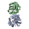
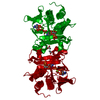
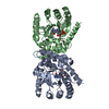
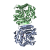
- Assembly
| Deposited unit | 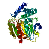
| ||||||||
|---|---|---|---|---|---|---|---|---|---|
| 1 | 
| ||||||||
| Unit cell |
| ||||||||
| Details | The second part of the biological assembly is generated by the two fold axis: -y+1/2, -x+1/2, -z+1/2. |
-Components
| #1: Protein | Mass: 33137.734 Da / Num. of mol.: 1 Source method: isolated from a genetically manipulated source Source: (gene. exp.) Janthinobacterium (bacteria) / Genus: Janthinobacterium / Strain: J3 / Gene: CarC / Plasmid: pECJ3 / Species (production host): Escherichia coli / Production host: References: UniProt: Q84II3, 2,6-dioxo-6-phenylhexa-3-enoate hydrolase |
|---|---|
| #2: Water | ChemComp-HOH /  Mass: 18.015 Da / Num. of mol.: 174 / Source method: isolated from a natural source / Formula: H2O Mass: 18.015 Da / Num. of mol.: 174 / Source method: isolated from a natural source / Formula: H2O |
-Experimental details
-Experiment
| Experiment | Method: X-RAY DIFFRACTION / Number of used crystals: 1 |
|---|
- Sample preparation
| Crystal | Density Matthews: 2.92 Å3/Da / Density % sol: 57.49 % | ||||||||||||||||||||||||
|---|---|---|---|---|---|---|---|---|---|---|---|---|---|---|---|---|---|---|---|---|---|---|---|---|---|
| Crystal grow | Temperature: 293 K / Method: vapor diffusion, hanging drop / pH: 5 Details: PEG 6000, citrate, pH 5.0, VAPOR DIFFUSION, HANGING DROP, temperature 293K | ||||||||||||||||||||||||
| Crystal grow | *PLUS Temperature: 20 ℃ / Method: vapor diffusion, hanging drop | ||||||||||||||||||||||||
| Components of the solutions | *PLUS
|
-Data collection
| Diffraction | Mean temperature: 100 K |
|---|---|
| Diffraction source | Source: ROTATING ANODE / Type: RIGAKU ULTRAX 18 / Wavelength: 1.5418 Å |
| Detector | Type: RIGAKU RAXIS IV++ / Detector: IMAGE PLATE / Date: Dec 14, 2000 / Details: mirrors |
| Radiation | Monochromator: YALE MIRRORS / Protocol: SINGLE WAVELENGTH / Monochromatic (M) / Laue (L): M / Scattering type: x-ray |
| Radiation wavelength | Wavelength: 1.5418 Å / Relative weight: 1 |
| Reflection | Resolution: 1.86→33.22 Å / Num. all: 30733 / Num. obs: 30496 / % possible obs: 99.2 % / Observed criterion σ(F): 0 / Observed criterion σ(I): 0 / Redundancy: 4.6 % / Biso Wilson estimate: 26.1 Å2 / Rmerge(I) obs: 0.073 / Net I/σ(I): 6.8 |
| Reflection shell | Resolution: 1.86→1.93 Å / Redundancy: 4.6 % / Rmerge(I) obs: 0.286 / Mean I/σ(I) obs: 2.4 / Num. unique all: 2978 / % possible all: 98.6 |
- Processing
| Software |
| ||||||||||||||||||||||||||||||||||||||||||||||||||||||||||||||||||||||||||||||||
|---|---|---|---|---|---|---|---|---|---|---|---|---|---|---|---|---|---|---|---|---|---|---|---|---|---|---|---|---|---|---|---|---|---|---|---|---|---|---|---|---|---|---|---|---|---|---|---|---|---|---|---|---|---|---|---|---|---|---|---|---|---|---|---|---|---|---|---|---|---|---|---|---|---|---|---|---|---|---|---|---|---|
| Refinement | Method to determine structure: MOLECULAR REPLACEMENT Starting model: PDB ENTRY 1IUP Resolution: 1.86→32.57 Å / Rfactor Rfree error: 0.006 / Isotropic thermal model: RESTRAINED / Cross valid method: THROUGHOUT / σ(F): 0 / Stereochemistry target values: Engh & Huber
| ||||||||||||||||||||||||||||||||||||||||||||||||||||||||||||||||||||||||||||||||
| Solvent computation | Solvent model: FLAT MODEL / Bsol: 49.2113 Å2 / ksol: 0.36267 e/Å3 | ||||||||||||||||||||||||||||||||||||||||||||||||||||||||||||||||||||||||||||||||
| Displacement parameters | Biso mean: 29.7 Å2
| ||||||||||||||||||||||||||||||||||||||||||||||||||||||||||||||||||||||||||||||||
| Refine analyze |
| ||||||||||||||||||||||||||||||||||||||||||||||||||||||||||||||||||||||||||||||||
| Refinement step | Cycle: LAST / Resolution: 1.86→32.57 Å
| ||||||||||||||||||||||||||||||||||||||||||||||||||||||||||||||||||||||||||||||||
| Refine LS restraints |
| ||||||||||||||||||||||||||||||||||||||||||||||||||||||||||||||||||||||||||||||||
| LS refinement shell | Resolution: 1.86→1.98 Å / Rfactor Rfree error: 0.018 / Total num. of bins used: 6
| ||||||||||||||||||||||||||||||||||||||||||||||||||||||||||||||||||||||||||||||||
| Xplor file |
| ||||||||||||||||||||||||||||||||||||||||||||||||||||||||||||||||||||||||||||||||
| Refinement | *PLUS % reflection Rfree: 5 % | ||||||||||||||||||||||||||||||||||||||||||||||||||||||||||||||||||||||||||||||||
| Solvent computation | *PLUS | ||||||||||||||||||||||||||||||||||||||||||||||||||||||||||||||||||||||||||||||||
| Displacement parameters | *PLUS | ||||||||||||||||||||||||||||||||||||||||||||||||||||||||||||||||||||||||||||||||
| Refine LS restraints | *PLUS
|
