 Movie
Movie Controller

[English] 日本語
 Yorodumi
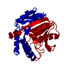
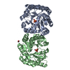
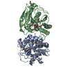
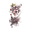
- PDB-6pky: Guinea pig N-acetylglucosamine-1-phosphodiester alpha-N-acetylglu... -
+ Open data

- Basic information
| Entry | Database: PDB / ID: 6pky | |||||||||
|---|---|---|---|---|---|---|---|---|---|---|
| Title | Guinea pig N-acetylglucosamine-1-phosphodiester alpha-N-acetylglucosaminidase (NAGPA) catalytic domain auto-inhibited by pro-peptide | |||||||||
 Components Components | N-acetylglucosamine-1-phosphodiester alpha-N-acetylglucosaminidase (NAGPA) | |||||||||
 Keywords Keywords | HYDROLASE / uncovering enzyme / mannose 6-phosphate / glycosidase / pro-peptide | |||||||||
| Function / homology | Phosphodiester glycosidase / Phosphodiester glycosidase / secretion of lysosomal enzymes / BROMIDE ION / N-acetylglucosamine-1-phosphodiester alpha-N-acetylglucosaminidase Function and homology information Function and homology information | |||||||||
| Biological species |  Cavia porcellus (domestic guinea pig) Cavia porcellus (domestic guinea pig) | |||||||||
| Method |  X-RAY DIFFRACTION / SYNCHROTRON / MOLECULAR REPLACEMENT / Resolution: 3 Å X-RAY DIFFRACTION / SYNCHROTRON / MOLECULAR REPLACEMENT / Resolution: 3 Å | |||||||||
 Authors Authors | Gorelik, A. / Illes, K. / Nagar, B. | |||||||||
| Funding support |  Canada, 1items Canada, 1items
| |||||||||
 Citation Citation | Journal: Structure / Year: 2020 Title: Crystal Structure of the Mannose-6-Phosphate Uncovering Enzyme. Authors: Gorelik, A. / Illes, K. / Nagar, B. | |||||||||
| History |
|
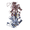
- Structure visualization
| Structure viewer | Molecule: MolmilJmol/JSmol |
|---|
- Downloads & links
-Download
| PDBx/mmCIF format | 6pky.cif.gz | 411.5 KB | Display | PDBx/mmCIF format |
|---|---|---|---|---|
| PDB format | pdb6pky.ent.gz | 340.2 KB | Display | PDB format |
| PDBx/mmJSON format | 6pky.json.gz | Tree view | PDBx/mmJSON format | |
| Others |  Other downloads Other downloads |
-Validation report
| Arichive directory | https://data.pdbj.org/pub/pdb/validation_reports/pk/6pkyftp://data.pdbj.org/pub/pdb/validation_reports/pk/6pky | HTTPS FTP |
|---|
-Related structure data
| Related structure data |  6pkgSC  6pkhC  6pkiC  6pkuC  6u11C S: Starting model for refinement C: citing same article ( |
|---|---|
| Similar structure data |


-Links
 PDBj
PDBj

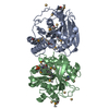
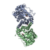
- Assembly
| Deposited unit | 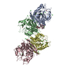
| ||||||||
|---|---|---|---|---|---|---|---|---|---|
| 1 | 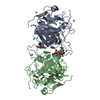
| ||||||||
| 2 | 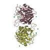
| ||||||||
| Unit cell |
|
-Components
-Protein , 1 types, 4 molecules ABCD
| #1: Protein | Mass: 33908.840 Da / Num. of mol.: 4 Source method: isolated from a genetically manipulated source Source: (gene. exp.) Cavia porcellus (domestic guinea pig) / Gene: NAGPA / Production host:   Spodoptera frugiperda (fall armyworm) / References: UniProt: H0VTT5 Spodoptera frugiperda (fall armyworm) / References: UniProt: H0VTT5 |
|---|
-Sugars , 2 types, 3 molecules 
| #2: Polysaccharide | 2-acetamido-2-deoxy-beta-D-glucopyranose-(1-4)-[alpha-L-fucopyranose-(1-6)]2-acetamido-2-deoxy-beta- ...2-acetamido-2-deoxy-beta-D-glucopyranose-(1-4)-[alpha-L-fucopyranose-(1-6)]2-acetamido-2-deoxy-beta-D-glucopyranose Source method: isolated from a genetically manipulated source |
|---|---|
| #3: Sugar |  Type: D-saccharide, beta linking / Mass: 221.208 Da / Num. of mol.: 2 Type: D-saccharide, beta linking / Mass: 221.208 Da / Num. of mol.: 2Source method: isolated from a genetically manipulated source Formula: C8H15NO6 |
-Non-polymers , 3 types, 155 molecules 


| #4: Chemical | ChemComp-BR /  Mass: 79.904 Da / Num. of mol.: 4 / Source method: obtained synthetically / Formula: Br Mass: 79.904 Da / Num. of mol.: 4 / Source method: obtained synthetically / Formula: Br#5: Chemical | ChemComp-CL /  Mass: 35.453 Da / Num. of mol.: 32 / Source method: obtained synthetically / Formula: Cl Mass: 35.453 Da / Num. of mol.: 32 / Source method: obtained synthetically / Formula: Cl#6: Water | ChemComp-HOH / | Mass: 18.015 Da / Num. of mol.: 119 / Source method: isolated from a natural source / Formula: H2O |
|---|
-Details
| Has ligand of interest | N |
|---|---|
| Has protein modification | Y |
-Experimental details
-Experiment
| Experiment | Method: X-RAY DIFFRACTION / Number of used crystals: 1 |
|---|
- Sample preparation
| Crystal | Density Matthews: 2.34 Å3/Da / Density % sol: 47.43 % |
|---|---|
| Crystal grow | Temperature: 295 K / Method: vapor diffusion, hanging drop / pH: 8.25 Details: 20% PEG 3350, 0.1M bis-tris propane pH 8.25, 0.2M NaBr |
-Data collection
| Diffraction | Mean temperature: 100 K / Serial crystal experiment: N |
|---|---|
| Diffraction source | Source: SYNCHROTRON / Site: ALS  / Beamline: 5.0.2 / Wavelength: 0.91977 Å / Beamline: 5.0.2 / Wavelength: 0.91977 Å |
| Detector | Type: DECTRIS PILATUS3 6M / Detector: PIXEL / Date: Dec 3, 2018 |
| Radiation | Protocol: SINGLE WAVELENGTH / Monochromatic (M) / Laue (L): M / Scattering type: x-ray |
| Radiation wavelength | Wavelength: 0.91977 Å / Relative weight: 1 |
| Reflection | Resolution: 3→50 Å / Num. obs: 24805 / % possible obs: 100 % / Redundancy: 23.2 % / Net I/σ(I): 10.6 |
| Reflection shell | Resolution: 3→3.11 Å / Num. unique obs: 2460 |
- Processing
| Software |
| ||||||||||||||||||||||||||||||||||||||||||||||||||||||||||||||||||||||||||||||||||||||||||||||||||||||||||||||||||||||||||||||||||||||||||||
|---|---|---|---|---|---|---|---|---|---|---|---|---|---|---|---|---|---|---|---|---|---|---|---|---|---|---|---|---|---|---|---|---|---|---|---|---|---|---|---|---|---|---|---|---|---|---|---|---|---|---|---|---|---|---|---|---|---|---|---|---|---|---|---|---|---|---|---|---|---|---|---|---|---|---|---|---|---|---|---|---|---|---|---|---|---|---|---|---|---|---|---|---|---|---|---|---|---|---|---|---|---|---|---|---|---|---|---|---|---|---|---|---|---|---|---|---|---|---|---|---|---|---|---|---|---|---|---|---|---|---|---|---|---|---|---|---|---|---|---|---|---|
| Refinement | Method to determine structure: MOLECULAR REPLACEMENT Starting model: 6PKG Resolution: 3→47.231 Å / Cross valid method: FREE R-VALUE / σ(F): 231.75 / Phase error: 29.54
| ||||||||||||||||||||||||||||||||||||||||||||||||||||||||||||||||||||||||||||||||||||||||||||||||||||||||||||||||||||||||||||||||||||||||||||
| Solvent computation | Shrinkage radii: 0.9 Å / VDW probe radii: 1.11 Å | ||||||||||||||||||||||||||||||||||||||||||||||||||||||||||||||||||||||||||||||||||||||||||||||||||||||||||||||||||||||||||||||||||||||||||||
| Refinement step | Cycle: LAST / Resolution: 3→47.231 Å
| ||||||||||||||||||||||||||||||||||||||||||||||||||||||||||||||||||||||||||||||||||||||||||||||||||||||||||||||||||||||||||||||||||||||||||||
| Refine LS restraints |
| ||||||||||||||||||||||||||||||||||||||||||||||||||||||||||||||||||||||||||||||||||||||||||||||||||||||||||||||||||||||||||||||||||||||||||||
| LS refinement shell |
|
