 Movie
Movie Controller

[English] 日本語
 Yorodumi
- PDB-4lyd: Crystal structure of the S105A mutant of a C-C hydrolase, DxnB2 f... -
+ Open data

- Basic information
| Entry | Database: PDB / ID: 4lyd | ||||||
|---|---|---|---|---|---|---|---|
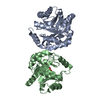
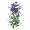
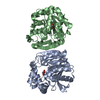
| Title | Crystal structure of the S105A mutant of a C-C hydrolase, DxnB2 from Sphingomonas wittichii RW1 | ||||||
 Components Components | MCP Hydrolase | ||||||
 Keywords Keywords | HYDROLASE / meta-cleavage product hydrolase / C-C bond hydrolase / alpha-beta hydrolase / dibenzo-p-dioxin degradation / dibenzofuran degradation | ||||||
| Function / homology | Alpha/beta hydrolase family / Alpha/beta hydrolase fold-1 / Alpha/Beta hydrolase fold, catalytic domain / Alpha/Beta hydrolase fold / hydrolase activity / Rossmann fold / 3-Layer(aba) Sandwich / Alpha Beta / Alpha/beta hydrolase fold Function and homology information Function and homology information | ||||||
| Biological species |  Sphingomonas wittichii (bacteria) Sphingomonas wittichii (bacteria) | ||||||
| Method |  X-RAY DIFFRACTION / SYNCHROTRON / MOLECULAR REPLACEMENT / molecular replacement / Resolution: 2.26 Å X-RAY DIFFRACTION / SYNCHROTRON / MOLECULAR REPLACEMENT / molecular replacement / Resolution: 2.26 Å | ||||||
 Authors Authors | Ghosh, S. / Bolin, J.T. / Bhowmik, S. | ||||||
 Citation Citation | Journal: Biochemistry / Year: 2013 Title: The Lid Domain of the MCP Hydrolase DxnB2 Contributes to the Reactivity toward Recalcitrant PCB Metabolites. Authors: Ruzzini, A.C. / Bhowmik, S. / Yam, K.C. / Ghosh, S. / Bolin, J.T. / Eltis, L.D. | ||||||
| History |
|
- Structure visualization
| Structure viewer | Molecule: MolmilJmol/JSmol |
|---|
- Downloads & links
-Download
| PDBx/mmCIF format | 4lyd.cif.gz | 64.5 KB | Display | PDBx/mmCIF format |
|---|---|---|---|---|
| PDB format | pdb4lyd.ent.gz | 47.6 KB | Display | PDB format |
| PDBx/mmJSON format | 4lyd.json.gz | Tree view | PDBx/mmJSON format | |
| Others |  Other downloads Other downloads |
-Validation report
| Arichive directory | https://data.pdbj.org/pub/pdb/validation_reports/ly/4lydftp://data.pdbj.org/pub/pdb/validation_reports/ly/4lyd | HTTPS FTP |
|---|
-Related structure data


-Links
 PDBj
PDBj

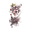
- Assembly
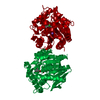
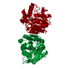
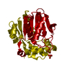
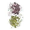
| Deposited unit | 
| ||||||||
|---|---|---|---|---|---|---|---|---|---|
| 1 | 
| ||||||||
| Unit cell |
|
-Components
| #1: Protein | Mass: 30208.531 Da / Num. of mol.: 1 / Mutation: S105A Source method: isolated from a genetically manipulated source Source: (gene. exp.) Sphingomonas wittichii (bacteria) / Strain: RW1 / Gene: dxnB2, Swit_3055 / Plasmid: pEMDXN1 / Production host: References: UniProt: A5VAT9, 2,6-dioxo-6-phenylhexa-3-enoate hydrolase |
|---|---|
| #2: Water | ChemComp-HOH /  Mass: 18.015 Da / Num. of mol.: 62 / Source method: isolated from a natural source / Formula: H2O Mass: 18.015 Da / Num. of mol.: 62 / Source method: isolated from a natural source / Formula: H2O |
-Experimental details
-Experiment
| Experiment | Method: X-RAY DIFFRACTION / Number of used crystals: 1 |
|---|
- Sample preparation
| Crystal | Density Matthews: 3.48 Å3/Da / Density % sol: 64.7 % |
|---|---|
| Crystal grow | Temperature: 298 K / Method: vapor diffusion, sitting drop / pH: 6.8 Details: 1.5 M sodium malonate, pH 6.8, VAPOR DIFFUSION, SITTING DROP, temperature 298K |
-Data collection
| Diffraction | Mean temperature: 100 K | |||||||||||||||||||||||||||||||||||||||||||||||||||||||||||||||||||||||||||||
|---|---|---|---|---|---|---|---|---|---|---|---|---|---|---|---|---|---|---|---|---|---|---|---|---|---|---|---|---|---|---|---|---|---|---|---|---|---|---|---|---|---|---|---|---|---|---|---|---|---|---|---|---|---|---|---|---|---|---|---|---|---|---|---|---|---|---|---|---|---|---|---|---|---|---|---|---|---|---|
| Diffraction source | Source: SYNCHROTRON / Site: APS  / Beamline: 23-ID-D / Wavelength: 1.033 Å / Beamline: 23-ID-D / Wavelength: 1.033 Å | |||||||||||||||||||||||||||||||||||||||||||||||||||||||||||||||||||||||||||||
| Detector | Type: MARMOSAIC 300 mm CCD / Detector: CCD / Date: Jul 1, 2010 | |||||||||||||||||||||||||||||||||||||||||||||||||||||||||||||||||||||||||||||
| Radiation | Monochromator: Si(111) / Protocol: SINGLE WAVELENGTH / Monochromatic (M) / Laue (L): M / Scattering type: x-ray | |||||||||||||||||||||||||||||||||||||||||||||||||||||||||||||||||||||||||||||
| Radiation wavelength | Wavelength: 1.033 Å / Relative weight: 1 | |||||||||||||||||||||||||||||||||||||||||||||||||||||||||||||||||||||||||||||
| Reflection | Resolution: 2.25→50 Å / Num. obs: 20422 / % possible obs: 93.5 % / Redundancy: 11.5 % / Rmerge(I) obs: 0.078 / Χ2: 1.206 / Net I/σ(I): 10.6 | |||||||||||||||||||||||||||||||||||||||||||||||||||||||||||||||||||||||||||||
| Reflection shell |
|
-Phasing
| Phasing | Method: molecular replacement | |||||||||
|---|---|---|---|---|---|---|---|---|---|---|
| Phasing MR | Rfactor: 39.22 / Model details: Phaser MODE: MR_AUTO
|
- Processing
| Software |
| |||||||||||||||||||||||||||||||||||||||||||||||||||||||||||||||||||||||||||
|---|---|---|---|---|---|---|---|---|---|---|---|---|---|---|---|---|---|---|---|---|---|---|---|---|---|---|---|---|---|---|---|---|---|---|---|---|---|---|---|---|---|---|---|---|---|---|---|---|---|---|---|---|---|---|---|---|---|---|---|---|---|---|---|---|---|---|---|---|---|---|---|---|---|---|---|---|
| Refinement | Method to determine structure: MOLECULAR REPLACEMENT / Resolution: 2.26→43.45 Å / Cor.coef. Fo:Fc: 0.951 / Cor.coef. Fo:Fc free: 0.928 / WRfactor Rfree: 0.2594 / WRfactor Rwork: 0.2047 / Occupancy max: 1 / Occupancy min: 1 / FOM work R set: 0.7796 / SU B: 7.053 / SU ML: 0.162 / SU R Cruickshank DPI: 0.2197 / SU Rfree: 0.2021 / Cross valid method: THROUGHOUT / σ(F): 0 / ESU R: 0.22 / ESU R Free: 0.202 / Stereochemistry target values: MAXIMUM LIKELIHOOD Details: HYDROGENS HAVE BEEN ADDED IN THE RIDING POSITIONS U VALUES : REFINED INDIVIDUALLY
| |||||||||||||||||||||||||||||||||||||||||||||||||||||||||||||||||||||||||||
| Solvent computation | Ion probe radii: 0.8 Å / Shrinkage radii: 0.8 Å / VDW probe radii: 1.2 Å / Solvent model: MASK | |||||||||||||||||||||||||||||||||||||||||||||||||||||||||||||||||||||||||||
| Displacement parameters | Biso max: 104.57 Å2 / Biso mean: 47.9948 Å2 / Biso min: 27.91 Å2
| |||||||||||||||||||||||||||||||||||||||||||||||||||||||||||||||||||||||||||
| Refinement step | Cycle: LAST / Resolution: 2.26→43.45 Å
| |||||||||||||||||||||||||||||||||||||||||||||||||||||||||||||||||||||||||||
| Refine LS restraints |
| |||||||||||||||||||||||||||||||||||||||||||||||||||||||||||||||||||||||||||
| LS refinement shell | Resolution: 2.256→2.315 Å / Total num. of bins used: 20
|
