 Movie
Movie Controller

[English] 日本語
 Yorodumi
- PDB-1uk6: Crystal structure of a meta-cleavage product hydrolase (CumD) com... -
+ Open data

- Basic information
| Entry | Database: PDB / ID: 1uk6 | ||||||
|---|---|---|---|---|---|---|---|
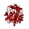
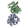
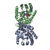
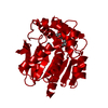
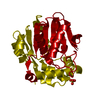
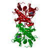
| Title | Crystal structure of a meta-cleavage product hydrolase (CumD) complexed with propionate | ||||||
 Components Components | 2-hydroxy-6-oxo-7-methylocta-2,4-dienoate hydrolase | ||||||
 Keywords Keywords | HYDROLASE / AROMATIC COMPOUNDS / CUMENE / ISOPROPYLBENZENE / META-CLEAVAGE COMPOUND HYDROLASE / POLYCHLORINATED BIPHENYL DEGRADATION / PSEUDOMONAS FLUORESCENS IP01 / ALPHA/BETA-HYDROLASE / SUBSTRATE SPECIFICITY / CUMENE DEGRADATION / PCB / BETA-KETOLASE | ||||||
| Function / homology |  Function and homology information Function and homology information | ||||||
| Biological species |  Pseudomonas fluorescens (bacteria) Pseudomonas fluorescens (bacteria) | ||||||
| Method |  X-RAY DIFFRACTION / SYNCHROTRON / FOURIER SYNTHESIS / Resolution: 1.95 Å X-RAY DIFFRACTION / SYNCHROTRON / FOURIER SYNTHESIS / Resolution: 1.95 Å | ||||||
 Authors Authors | Fushinobu, S. / Jun, S.-Y. / Hidaka, M. / Nojiri, H. / Yamane, H. / Shoun, H. / Omori, T. / Wakagi, T. | ||||||
 Citation Citation | Journal: BIOSCI.BIOTECHNOL.BIOCHEM. / Year: 2005 Title: A Series of Crystal Structures of a meta-Cleavage Product Hydrolase from Pseudomonas fluorescens IP01 (CumD) Complexed with Various Cleavage Products Authors: Fushinobu, S. / Jun, S.-Y. / Hidaka, M. / Nojiri, H. / Yamane, H. / Shoun, H. / Omori, T. / Wakagi, T. | ||||||
| History |
|
- Structure visualization
| Structure viewer | Molecule: MolmilJmol/JSmol |
|---|
- Downloads & links
-Download
| PDBx/mmCIF format | 1uk6.cif.gz | 75.3 KB | Display | PDBx/mmCIF format |
|---|---|---|---|---|
| PDB format | pdb1uk6.ent.gz | 54.6 KB | Display | PDB format |
| PDBx/mmJSON format | 1uk6.json.gz | Tree view | PDBx/mmJSON format | |
| Others |  Other downloads Other downloads |
-Validation report
| Arichive directory | https://data.pdbj.org/pub/pdb/validation_reports/uk/1uk6ftp://data.pdbj.org/pub/pdb/validation_reports/uk/1uk6 | HTTPS FTP |
|---|
-Related structure data
| Related structure data | 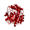 1uk7C  1uk8C  1uk9C  1ukaC 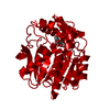 1ukbC  1iupS S: Starting model for refinement C: citing same article ( |
|---|---|
| Similar structure data |


-Links
 PDBj
PDBj


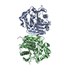
- Assembly
| Deposited unit | 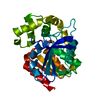
| ||||||||
|---|---|---|---|---|---|---|---|---|---|
| 1 | 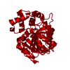
| ||||||||
| Unit cell |
| ||||||||
| Details | The second part of the biological assembly is generated by the two fold axis: x, -y+1, -z. |
-Components
| #1: Protein | Mass: 31506.607 Da / Num. of mol.: 1 / Mutation: S103A Source method: isolated from a genetically manipulated source Source: (gene. exp.) Pseudomonas fluorescens (bacteria) / Gene: CUMD / Plasmid: PIP140 / Production host: | ||
|---|---|---|---|
| #2: Chemical |   Mass: 74.079 Da / Num. of mol.: 2 / Source method: obtained synthetically / Formula: C3H6O2 Mass: 74.079 Da / Num. of mol.: 2 / Source method: obtained synthetically / Formula: C3H6O2#3: Water | ChemComp-HOH / |  Mass: 18.015 Da / Num. of mol.: 334 / Source method: isolated from a natural source / Formula: H2O Mass: 18.015 Da / Num. of mol.: 334 / Source method: isolated from a natural source / Formula: H2O |
-Experimental details
-Experiment
| Experiment | Method: X-RAY DIFFRACTION / Number of used crystals: 1 |
|---|
- Sample preparation
| Crystal | Density Matthews: 2.53 Å3/Da / Density % sol: 51.01 % |
|---|---|
| Crystal grow | Temperature: 283 K / Method: vapor diffusion, hanging drop / pH: 5.1 Details: PEG4000, AMMONIUM propionate, SODIUM propionate, pH 5.1, VAPOR DIFFUSION, HANGING DROP, temperature 283K |
-Data collection
| Diffraction | Mean temperature: 100 K |
|---|---|
| Diffraction source | Source: SYNCHROTRON / Site: Photon Factory  / Beamline: BL-6A / Wavelength: 0.978 Å / Beamline: BL-6A / Wavelength: 0.978 Å |
| Detector | Type: ADSC QUANTUM 4 / Detector: CCD / Date: Apr 16, 2002 |
| Radiation | Monochromator: Si 111 / Protocol: SINGLE WAVELENGTH / Monochromatic (M) / Laue (L): M / Scattering type: x-ray |
| Radiation wavelength | Wavelength: 0.978 Å / Relative weight: 1 |
| Reflection | Resolution: 1.95→27.46 Å / Num. all: 26055 / Num. obs: 26055 / % possible obs: 99.9 % / Observed criterion σ(F): 0 / Observed criterion σ(I): 0 / Redundancy: 3.6 % / Biso Wilson estimate: 5.8 Å2 / Rsym value: 0.115 / Net I/σ(I): 6.3 |
| Reflection shell | Resolution: 1.95→2.06 Å / Mean I/σ(I) obs: 2.1 / Rsym value: 0.348 / % possible all: 100 |
- Processing
| Software |
| ||||||||||||||||||||||||||||||||||||||||||||||||||||||||||||||||||||||||||||||||
|---|---|---|---|---|---|---|---|---|---|---|---|---|---|---|---|---|---|---|---|---|---|---|---|---|---|---|---|---|---|---|---|---|---|---|---|---|---|---|---|---|---|---|---|---|---|---|---|---|---|---|---|---|---|---|---|---|---|---|---|---|---|---|---|---|---|---|---|---|---|---|---|---|---|---|---|---|---|---|---|---|---|
| Refinement | Method to determine structure: FOURIER SYNTHESIS Starting model: PDB ENTRY 1IUP Resolution: 1.95→27.42 Å / Rfactor Rfree error: 0.006 / Data cutoff high absF: 2243410.22 / Data cutoff low absF: 0 / Isotropic thermal model: RESTRAINED / Cross valid method: THROUGHOUT / σ(F): 0 / Stereochemistry target values: Engh & Huber
| ||||||||||||||||||||||||||||||||||||||||||||||||||||||||||||||||||||||||||||||||
| Solvent computation | Solvent model: FLAT MODEL / Bsol: 50.2379 Å2 / ksol: 0.337363 e/Å3 | ||||||||||||||||||||||||||||||||||||||||||||||||||||||||||||||||||||||||||||||||
| Displacement parameters | Biso mean: 12.9 Å2
| ||||||||||||||||||||||||||||||||||||||||||||||||||||||||||||||||||||||||||||||||
| Refine analyze |
| ||||||||||||||||||||||||||||||||||||||||||||||||||||||||||||||||||||||||||||||||
| Refinement step | Cycle: LAST / Resolution: 1.95→27.42 Å
| ||||||||||||||||||||||||||||||||||||||||||||||||||||||||||||||||||||||||||||||||
| Refine LS restraints |
| ||||||||||||||||||||||||||||||||||||||||||||||||||||||||||||||||||||||||||||||||
| LS refinement shell | Resolution: 1.95→2.07 Å / Rfactor Rfree error: 0.016 / Total num. of bins used: 6
| ||||||||||||||||||||||||||||||||||||||||||||||||||||||||||||||||||||||||||||||||
| Xplor file |
|
