 Movie
Movie Controller

[English] 日本語
 Yorodumi
- PDB-4ekd: Structure of human regulator of G protein signaling 2 (RGS2) in c... -
+ Open data

- Basic information
| Entry | Database: PDB / ID: 4ekd | ||||||
|---|---|---|---|---|---|---|---|
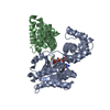
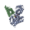
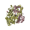
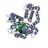
| Title | Structure of human regulator of G protein signaling 2 (RGS2) in complex with murine Galpha-q(R183C) | ||||||
 Components Components |
| ||||||
 Keywords Keywords | SIGNALING PROTEIN/INHIBITOR / GTP-binding / Regulator of G protein signaling / homology domain / GTPase activation / SIGNALING PROTEIN-INHIBITOR complex | ||||||
| Function / homology |  Function and homology information Function and homology informationregulation of adenylate cyclase-inhibiting adrenergic receptor signaling pathway / negative regulation of glycine import across plasma membrane / Fatty Acids bound to GPR40 (FFAR1) regulate insulin secretion / PLC beta mediated events / Acetylcholine regulates insulin secretion / forebrain neuron development / regulation of melanocyte differentiation / Cooperation of PDCL (PhLP1) and TRiC/CCT in G-protein beta folding / Thromboxane signalling through TP receptor / Thrombin signalling through proteinase activated receptors (PARs) ...regulation of adenylate cyclase-inhibiting adrenergic receptor signaling pathway / negative regulation of glycine import across plasma membrane / Fatty Acids bound to GPR40 (FFAR1) regulate insulin secretion / PLC beta mediated events / Acetylcholine regulates insulin secretion / forebrain neuron development / regulation of melanocyte differentiation / Cooperation of PDCL (PhLP1) and TRiC/CCT in G-protein beta folding / Thromboxane signalling through TP receptor / Thrombin signalling through proteinase activated receptors (PARs) / adenylate cyclase-activating G protein-coupled cAMP receptor signaling pathway / Turbulent (oscillatory, disturbed) flow shear stress activates signaling by PIEZO1 and integrins in endothelial cells / G-protein activation / G alpha (q) signalling events / phospholipase C-activating G protein-coupled acetylcholine receptor signaling pathway / endothelin receptor signaling pathway / developmental pigmentation / phospholipase C-activating G protein-coupled glutamate receptor signaling pathway / phospholipase C-activating dopamine receptor signaling pathway / negative regulation of cardiac muscle hypertrophy / negative regulation of cell growth involved in cardiac muscle cell development / ADP signalling through P2Y purinoceptor 1 / High laminar flow shear stress activates signaling by PIEZO1 and PECAM1:CDH5:KDR in endothelial cells / phospholipase C-activating serotonin receptor signaling pathway / regulation of platelet activation / cranial skeletal system development / relaxation of vascular associated smooth muscle / regulation of G protein-coupled receptor signaling pathway / maternal behavior / regulation of canonical Wnt signaling pathway / relaxation of cardiac muscle / negative regulation of JNK cascade / embryonic digit morphogenesis / negative regulation of G protein-coupled receptor signaling pathway / glutamate receptor signaling pathway / positive regulation of phospholipase C-activating G protein-coupled receptor signaling pathway / neuron remodeling / ligand-gated ion channel signaling pathway / alkylglycerophosphoethanolamine phosphodiesterase activity / negative regulation of potassium ion transport / maternal process involved in female pregnancy / action potential / G-protein alpha-subunit binding / postsynaptic cytosol / beta-tubulin binding / cellular response to acidic pH / enzyme regulator activity / adenylate cyclase inhibitor activity / hormone-mediated signaling pathway / positive regulation of cardiac muscle contraction / brown fat cell differentiation / GTPase activator activity / post-embryonic development / response to amphetamine / skeletal system development / neuropeptide signaling pathway / response to prostaglandin E / positive regulation of neuron projection development / caveola / G protein-coupled receptor binding / regulation of blood pressure / cytoplasmic side of plasma membrane / G-protein beta/gamma-subunit complex binding / positive regulation of insulin secretion / heterotrimeric G-protein complex / heart development / presynapse / adenylate cyclase-activating G protein-coupled receptor signaling pathway / cell body / G protein activity / spermatogenesis / nuclear membrane / G alpha (q) signalling events / phospholipase C-activating G protein-coupled receptor signaling pathway / Hydrolases; Acting on acid anhydrides; Acting on GTP to facilitate cellular and subcellular movement / response to ethanol / calmodulin binding / negative regulation of translation / protein stabilization / G protein-coupled receptor signaling pathway / GTPase activity / dendrite / negative regulation of apoptotic process / GTP binding / protein-containing complex binding / nucleolus / Golgi apparatus / mitochondrion / membrane / metal ion binding / nucleus / plasma membrane / cytoplasm / cytosol Similarity search - Function | ||||||
| Biological species |   Homo sapiens (human) Homo sapiens (human) | ||||||
| Method |  X-RAY DIFFRACTION / SYNCHROTRON / MOLECULAR REPLACEMENT / Resolution: 2.71 Å X-RAY DIFFRACTION / SYNCHROTRON / MOLECULAR REPLACEMENT / Resolution: 2.71 Å | ||||||
 Authors Authors | Tesmer, J.J.G. / Nance, M.R. | ||||||
 Citation Citation | Journal: Structure / Year: 2013 Title: Structural and functional analysis of the regulator of G protein signaling 2-g alpha q complex. Authors: Nance, M.R. / Kreutz, B. / Tesmer, V.M. / Sterne-Marr, R. / Kozasa, T. / Tesmer, J.J. | ||||||
| History |
|
- Structure visualization
| Structure viewer | Molecule: MolmilJmol/JSmol |
|---|
- Downloads & links
-Download
| PDBx/mmCIF format | 4ekd.cif.gz | 206.9 KB | Display | PDBx/mmCIF format |
|---|---|---|---|---|
| PDB format | pdb4ekd.ent.gz | 165.1 KB | Display | PDB format |
| PDBx/mmJSON format | 4ekd.json.gz | Tree view | PDBx/mmJSON format | |
| Others |  Other downloads Other downloads |
-Validation report
| Arichive directory | https://data.pdbj.org/pub/pdb/validation_reports/ek/4ekdftp://data.pdbj.org/pub/pdb/validation_reports/ek/4ekd | HTTPS FTP |
|---|
-Related structure data
| Related structure data |  4ekcC  2af0S 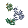 2bcjS S: Starting model for refinement C: citing same article ( |
|---|---|
| Similar structure data |
-Links
 PDBj
PDBj


- Assembly
| Deposited unit | 
| ||||||||
|---|---|---|---|---|---|---|---|---|---|
| 1 |
| ||||||||
| Unit cell |
|
-Components
-Protein , 2 types, 2 molecules AB
| #1: Protein | Mass: 40634.172 Da / Num. of mol.: 1 / Fragment: UNP residues 18-359 / Mutation: E125D, N126V, Y128D, V129Y, D130A, R183C Source method: isolated from a genetically manipulated source Source: (gene. exp.)  Trichoplusia ni (cabbage looper) / References: UniProt: P21279, EC: 3.6.5.1 Trichoplusia ni (cabbage looper) / References: UniProt: P21279, EC: 3.6.5.1 |
|---|---|
| #2: Protein | Mass: 15871.833 Da / Num. of mol.: 1 / Fragment: RGS domain, UNP residues 72-203 Source method: isolated from a genetically manipulated source Source: (gene. exp.) Homo sapiens (human) / Gene: RGS2, G0S8, GIG31 / Production host:  |
-Non-polymers , 7 types, 24 molecules 


| #3: Chemical | ChemComp-GDP /  Type: RNA linking / Mass: 443.201 Da / Num. of mol.: 1 / Source method: obtained synthetically / Formula: C10H15N5O11P2 / Comment: GDP, energy-carrying molecule*YM Type: RNA linking / Mass: 443.201 Da / Num. of mol.: 1 / Source method: obtained synthetically / Formula: C10H15N5O11P2 / Comment: GDP, energy-carrying molecule*YM | ||||||||
|---|---|---|---|---|---|---|---|---|---|
| #4: Chemical | ChemComp-ALF /  Mass: 102.975 Da / Num. of mol.: 1 / Source method: obtained synthetically / Formula: AlF4 Mass: 102.975 Da / Num. of mol.: 1 / Source method: obtained synthetically / Formula: AlF4 | ||||||||
| #5: Chemical |  Mass: 58.933 Da / Num. of mol.: 2 / Source method: obtained synthetically / Formula: Co Mass: 58.933 Da / Num. of mol.: 2 / Source method: obtained synthetically / Formula: Co#6: Chemical |  Mass: 35.453 Da / Num. of mol.: 2 / Source method: obtained synthetically / Formula: Cl Mass: 35.453 Da / Num. of mol.: 2 / Source method: obtained synthetically / Formula: Cl#7: Chemical | ChemComp-MG / |  Mass: 24.305 Da / Num. of mol.: 1 / Source method: obtained synthetically / Formula: Mg Mass: 24.305 Da / Num. of mol.: 1 / Source method: obtained synthetically / Formula: Mg#8: Chemical | ChemComp-MES / |  Mass: 195.237 Da / Num. of mol.: 1 / Source method: obtained synthetically / Formula: C6H13NO4S / Comment: pH buffer*YM Mass: 195.237 Da / Num. of mol.: 1 / Source method: obtained synthetically / Formula: C6H13NO4S / Comment: pH buffer*YM#9: Water | ChemComp-HOH / | Mass: 18.015 Da / Num. of mol.: 16 / Source method: isolated from a natural source / Formula: H2O |
-Experimental details
-Experiment
| Experiment | Method: X-RAY DIFFRACTION / Number of used crystals: 1 |
|---|
- Sample preparation
| Crystal | Density Matthews: 2.77 Å3/Da / Density % sol: 55.67 % |
|---|---|
| Crystal grow | Temperature: 277 K / Method: vapor diffusion, hanging drop / pH: 5.5 Details: 12% PEG 8,000, 15 mM CoCl2, 200 mM NaCl, and 100 mM MES 5.5, VAPOR DIFFUSION, HANGING DROP, temperature 277K |
-Data collection
| Diffraction | Mean temperature: 110 K |
|---|---|
| Diffraction source | Source: SYNCHROTRON / Site: APS  / Beamline: 21-ID-D / Wavelength: 1.0782 Å / Beamline: 21-ID-D / Wavelength: 1.0782 Å |
| Detector | Type: RAYONIX MX-300 / Detector: CCD / Date: Oct 13, 2010 / Details: KB Bimorph Mirrors |
| Radiation | Monochromator: Si(111) double crystal / Protocol: SINGLE WAVELENGTH / Monochromatic (M) / Laue (L): M / Scattering type: x-ray |
| Radiation wavelength | Wavelength: 1.0782 Å / Relative weight: 1 |
| Reflection | Resolution: 2.7→30 Å / Num. all: 12037 / Num. obs: 11782 / % possible obs: 64 % / Observed criterion σ(I): -3 / Redundancy: 4 % / Rsym value: 0.065 / Net I/σ(I): 24.9 |
| Reflection shell | Resolution: 2.7→2.75 Å / Redundancy: 2.7 % / Mean I/σ(I) obs: 1.6 / Num. unique all: 189 / Rsym value: 0.441 / % possible all: 21.4 |
- Processing
| Software |
| ||||||||||||||||||||||||||||||||||||||||||||||||||||||||||||||||||||||||||||||||||||||||||||||||||||||||||||||||||||||||||||||||||||||||||||||||||||||||||||||||||||||||||
|---|---|---|---|---|---|---|---|---|---|---|---|---|---|---|---|---|---|---|---|---|---|---|---|---|---|---|---|---|---|---|---|---|---|---|---|---|---|---|---|---|---|---|---|---|---|---|---|---|---|---|---|---|---|---|---|---|---|---|---|---|---|---|---|---|---|---|---|---|---|---|---|---|---|---|---|---|---|---|---|---|---|---|---|---|---|---|---|---|---|---|---|---|---|---|---|---|---|---|---|---|---|---|---|---|---|---|---|---|---|---|---|---|---|---|---|---|---|---|---|---|---|---|---|---|---|---|---|---|---|---|---|---|---|---|---|---|---|---|---|---|---|---|---|---|---|---|---|---|---|---|---|---|---|---|---|---|---|---|---|---|---|---|---|---|---|---|---|---|---|---|---|
| Refinement | Method to determine structure: MOLECULAR REPLACEMENT Starting model: PDB entries 2AF0 and 2BCJ Resolution: 2.71→29.98 Å / Cor.coef. Fo:Fc: 0.967 / Cor.coef. Fo:Fc free: 0.952 / SU B: 44.27 / SU ML: 0.342 / Cross valid method: THROUGHOUT / ESU R Free: 0.471 / Stereochemistry target values: MAXIMUM LIKELIHOOD / Details: HYDROGENS HAVE BEEN ADDED IN THE RIDING POSITIONS
| ||||||||||||||||||||||||||||||||||||||||||||||||||||||||||||||||||||||||||||||||||||||||||||||||||||||||||||||||||||||||||||||||||||||||||||||||||||||||||||||||||||||||||
| Solvent computation | Ion probe radii: 0.8 Å / Shrinkage radii: 0.8 Å / VDW probe radii: 1.2 Å / Solvent model: MASK | ||||||||||||||||||||||||||||||||||||||||||||||||||||||||||||||||||||||||||||||||||||||||||||||||||||||||||||||||||||||||||||||||||||||||||||||||||||||||||||||||||||||||||
| Displacement parameters | Biso mean: 96.352 Å2
| ||||||||||||||||||||||||||||||||||||||||||||||||||||||||||||||||||||||||||||||||||||||||||||||||||||||||||||||||||||||||||||||||||||||||||||||||||||||||||||||||||||||||||
| Refinement step | Cycle: LAST / Resolution: 2.71→29.98 Å
| ||||||||||||||||||||||||||||||||||||||||||||||||||||||||||||||||||||||||||||||||||||||||||||||||||||||||||||||||||||||||||||||||||||||||||||||||||||||||||||||||||||||||||
| Refine LS restraints |
| ||||||||||||||||||||||||||||||||||||||||||||||||||||||||||||||||||||||||||||||||||||||||||||||||||||||||||||||||||||||||||||||||||||||||||||||||||||||||||||||||||||||||||
| LS refinement shell | Resolution: 2.71→2.782 Å / Total num. of bins used: 20
| ||||||||||||||||||||||||||||||||||||||||||||||||||||||||||||||||||||||||||||||||||||||||||||||||||||||||||||||||||||||||||||||||||||||||||||||||||||||||||||||||||||||||||
| Refinement TLS params. | Method: refined / Refine-ID: X-RAY DIFFRACTION
| ||||||||||||||||||||||||||||||||||||||||||||||||||||||||||||||||||||||||||||||||||||||||||||||||||||||||||||||||||||||||||||||||||||||||||||||||||||||||||||||||||||||||||
| Refinement TLS group |
|
