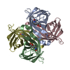
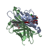
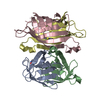
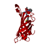
 Movie
Movie Controller


+ Open data

- Basic information
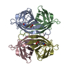
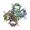
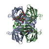
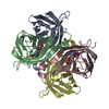
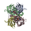
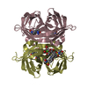
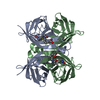
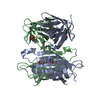
| Entry | Database: PDB / ID: 4bx5 | ||||||
|---|---|---|---|---|---|---|---|
| Title | cis-divalent streptavidin | ||||||
 Components Components | (STREPTAVIDIN) x 2 | ||||||
 Keywords Keywords | BIOTIN-BINDING PROTEIN / BIOTIN / AVIDIN | ||||||
| Function / homology |  Function and homology information Function and homology information | ||||||
| Biological species |  STREPTOMYCES AVIDINII (bacteria) STREPTOMYCES AVIDINII (bacteria) | ||||||
| Method |  X-RAY DIFFRACTION / SYNCHROTRON / MOLECULAR REPLACEMENT / Resolution: 1.431 Å X-RAY DIFFRACTION / SYNCHROTRON / MOLECULAR REPLACEMENT / Resolution: 1.431 Å | ||||||
 Authors Authors | Fairhead, M. / Krndija, D. / Lowe, E.D. / Howarth, M. | ||||||
 Citation Citation | Journal: J.Mol.Biol. / Year: 2014 Title: Plug-and-Play Pairing Via Defined Divalent Streptavidins. Authors: Fairhead, M. / Krndija, D. / Lowe, E.D. / Howarth, M. | ||||||
| History |
| ||||||
| Remark 700 | SHEET DETERMINATION METHOD: DSSP THE SHEETS PRESENTED AS "AA" IN EACH CHAIN ON SHEET RECORDS BELOW ... SHEET DETERMINATION METHOD: DSSP THE SHEETS PRESENTED AS "AA" IN EACH CHAIN ON SHEET RECORDS BELOW IS ACTUALLY AN 8-STRANDED BARREL THIS IS REPRESENTED BY A 9-STRANDED SHEET IN WHICH THE FIRST AND LAST STRANDS ARE IDENTICAL. THE SHEETS PRESENTED AS "BA" IN EACH CHAIN ON SHEET RECORDS BELOW IS ACTUALLY AN 8-STRANDED BARREL THIS IS REPRESENTED BY A 9-STRANDED SHEET IN WHICH THE FIRST AND LAST STRANDS ARE IDENTICAL. THE SHEETS PRESENTED AS "CA" IN EACH CHAIN ON SHEET RECORDS BELOW IS ACTUALLY AN 8-STRANDED BARREL THIS IS REPRESENTED BY A 9-STRANDED SHEET IN WHICH THE FIRST AND LAST STRANDS ARE IDENTICAL. THE SHEETS PRESENTED AS "DA" IN EACH CHAIN ON SHEET RECORDS BELOW IS ACTUALLY AN 8-STRANDED BARREL THIS IS REPRESENTED BY A 9-STRANDED SHEET IN WHICH THE FIRST AND LAST STRANDS ARE IDENTICAL. |
- Structure visualization
| Structure viewer | Molecule: MolmilJmol/JSmol |
|---|
- Downloads & links
-Download
| PDBx/mmCIF format | 4bx5.cif.gz | 118.1 KB | Display | PDBx/mmCIF format |
|---|---|---|---|---|
| PDB format | pdb4bx5.ent.gz | 90.9 KB | Display | PDB format |
| PDBx/mmJSON format | 4bx5.json.gz | Tree view | PDBx/mmJSON format | |
| Others |  Other downloads Other downloads |
-Validation report
| Arichive directory | https://data.pdbj.org/pub/pdb/validation_reports/bx/4bx5ftp://data.pdbj.org/pub/pdb/validation_reports/bx/4bx5 | HTTPS FTP |
|---|
-Related structure data
| Related structure data |  4bx6C  4bx7C  3ry1S C: citing same article ( S: Starting model for refinement |
|---|---|
| Similar structure data |

-Links
 PDBj
PDBj- Assembly
| Deposited unit | 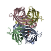
| ||||||||
|---|---|---|---|---|---|---|---|---|---|
| 1 |
| ||||||||
| Unit cell |
|
-Components
| #1: Protein | Mass: 14343.168 Da / Num. of mol.: 2 / Fragment: RESIDUES 37-163 / Mutation: YES Source method: isolated from a genetically manipulated source Source: (gene. exp.) STREPTOMYCES AVIDINII (bacteria) / Production host: #2: Protein | Mass: 13281.336 Da / Num. of mol.: 2 / Fragment: RESIDUES 37-163 Source method: isolated from a genetically manipulated source Source: (gene. exp.) STREPTOMYCES AVIDINII (bacteria) / Production host: #3: Chemical | ChemComp-EDO /   Mass: 62.068 Da / Num. of mol.: 4 / Source method: obtained synthetically / Formula: C2H6O2 Mass: 62.068 Da / Num. of mol.: 4 / Source method: obtained synthetically / Formula: C2H6O2#4: Chemical |   Mass: 194.226 Da / Num. of mol.: 2 / Source method: obtained synthetically / Formula: C8H18O5 / Comment: precipitant*YM Mass: 194.226 Da / Num. of mol.: 2 / Source method: obtained synthetically / Formula: C8H18O5 / Comment: precipitant*YM#5: Water | ChemComp-HOH / |  Mass: 18.015 Da / Num. of mol.: 517 / Source method: isolated from a natural source / Formula: H2O Mass: 18.015 Da / Num. of mol.: 517 / Source method: isolated from a natural source / Formula: H2O |
|---|
-Experimental details
-Experiment
| Experiment | Method: X-RAY DIFFRACTION / Number of used crystals: 1 |
|---|
- Sample preparation
| Crystal | Density Matthews: 2.12 Å3/Da / Density % sol: 41.44 % / Description: NONE |
|---|---|
| Crystal grow | pH: 6.5 Details: 2.5 % W/V POLYETHYLENE GLYCOL (PEG) 1000, 12.5 % W/V PEG 3350, 12.5 % V/V MPD, 30 MM OF ETHYLENE GLYCOL MIX (DI-ETHYLENEGLYCOL, TRI-ETHYLENEGLYCOL, TETRA-ETHYLENEGLYCOL, PENTA-ETHYLENEGLYCOL) ...Details: 2.5 % W/V POLYETHYLENE GLYCOL (PEG) 1000, 12.5 % W/V PEG 3350, 12.5 % V/V MPD, 30 MM OF ETHYLENE GLYCOL MIX (DI-ETHYLENEGLYCOL, TRI-ETHYLENEGLYCOL, TETRA-ETHYLENEGLYCOL, PENTA-ETHYLENEGLYCOL) AND 0.1 M MES/IMIDAZOLE PH 6.5, CORRESPONDING TO CONDITION E4 OF THE MORPHEUS SCREEN |
-Data collection
| Diffraction | Mean temperature: 100 K |
|---|---|
| Diffraction source | Source: SYNCHROTRON / Site: Diamond  / Beamline: I04-1 / Wavelength: 0.9173 / Beamline: I04-1 / Wavelength: 0.9173 |
| Detector | Type: MARRESEARCH 300MM / Detector: CCD / Date: Mar 9, 2012 |
| Radiation | Protocol: SINGLE WAVELENGTH / Monochromatic (M) / Laue (L): M / Scattering type: x-ray |
| Radiation wavelength | Wavelength: 0.9173 Å / Relative weight: 1 |
| Reflection | Resolution: 1.43→42.12 Å / Num. obs: 75108 / % possible obs: 92.6 % / Observed criterion σ(I): 2 / Redundancy: 3.6 % / Biso Wilson estimate: 14.65 Å2 / Rmerge(I) obs: 0.06 / Net I/σ(I): 11.4 |
| Reflection shell | Resolution: 1.43→1.48 Å / Redundancy: 3.6 % / Rmerge(I) obs: 0.58 / Mean I/σ(I) obs: 2.21 / % possible all: 91.6 |
- Processing
| Software |
| ||||||||||||||||||||||||||||||||||||||||||||||||||||||||||||||||||||||||||||||||||||||||||||||||||||||||||||||||||||||||||||||||||||||||||||||||||||||||||||||||||||||||||||||||||||||||||||||||||||
|---|---|---|---|---|---|---|---|---|---|---|---|---|---|---|---|---|---|---|---|---|---|---|---|---|---|---|---|---|---|---|---|---|---|---|---|---|---|---|---|---|---|---|---|---|---|---|---|---|---|---|---|---|---|---|---|---|---|---|---|---|---|---|---|---|---|---|---|---|---|---|---|---|---|---|---|---|---|---|---|---|---|---|---|---|---|---|---|---|---|---|---|---|---|---|---|---|---|---|---|---|---|---|---|---|---|---|---|---|---|---|---|---|---|---|---|---|---|---|---|---|---|---|---|---|---|---|---|---|---|---|---|---|---|---|---|---|---|---|---|---|---|---|---|---|---|---|---|---|---|---|---|---|---|---|---|---|---|---|---|---|---|---|---|---|---|---|---|---|---|---|---|---|---|---|---|---|---|---|---|---|---|---|---|---|---|---|---|---|---|---|---|---|---|---|---|---|---|
| Refinement | Method to determine structure: MOLECULAR REPLACEMENT Starting model: PDB ENTRY 3RY1 Resolution: 1.431→42.12 Å / SU ML: 0.14 / σ(F): 1.35 / Phase error: 19.95 / Stereochemistry target values: ML
| ||||||||||||||||||||||||||||||||||||||||||||||||||||||||||||||||||||||||||||||||||||||||||||||||||||||||||||||||||||||||||||||||||||||||||||||||||||||||||||||||||||||||||||||||||||||||||||||||||||
| Solvent computation | Shrinkage radii: 0.9 Å / VDW probe radii: 1.11 Å / Solvent model: FLAT BULK SOLVENT MODEL | ||||||||||||||||||||||||||||||||||||||||||||||||||||||||||||||||||||||||||||||||||||||||||||||||||||||||||||||||||||||||||||||||||||||||||||||||||||||||||||||||||||||||||||||||||||||||||||||||||||
| Displacement parameters | Biso mean: 22 Å2 | ||||||||||||||||||||||||||||||||||||||||||||||||||||||||||||||||||||||||||||||||||||||||||||||||||||||||||||||||||||||||||||||||||||||||||||||||||||||||||||||||||||||||||||||||||||||||||||||||||||
| Refinement step | Cycle: LAST / Resolution: 1.431→42.12 Å
| ||||||||||||||||||||||||||||||||||||||||||||||||||||||||||||||||||||||||||||||||||||||||||||||||||||||||||||||||||||||||||||||||||||||||||||||||||||||||||||||||||||||||||||||||||||||||||||||||||||
| Refine LS restraints |
| ||||||||||||||||||||||||||||||||||||||||||||||||||||||||||||||||||||||||||||||||||||||||||||||||||||||||||||||||||||||||||||||||||||||||||||||||||||||||||||||||||||||||||||||||||||||||||||||||||||
| LS refinement shell |
|
