 Movie
Movie Controller

[English] 日本語
 Yorodumi
- PDB-1mep: Crystal Structure of Streptavidin Double Mutant S45A/D128A with B... -
+ Open data

- Basic information
| Entry | Database: PDB / ID: 1mep | ||||||
|---|---|---|---|---|---|---|---|
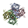
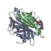
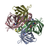
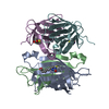
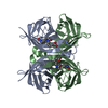
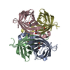
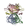
| Title | Crystal Structure of Streptavidin Double Mutant S45A/D128A with Biotin: Cooperative Hydrogen-Bond Interactions in the Streptavidin-Biotin System. | ||||||
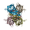
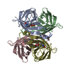
 Components Components | Streptavidin | ||||||
 Keywords Keywords | Biotin-binding protein / homotetramer | ||||||
| Function / homology |  Function and homology information Function and homology information | ||||||
| Biological species |  Streptomyces avidinii (bacteria) Streptomyces avidinii (bacteria) | ||||||
| Method |  X-RAY DIFFRACTION / SYNCHROTRON / ISOMORPHOUS / Resolution: 1.65 Å X-RAY DIFFRACTION / SYNCHROTRON / ISOMORPHOUS / Resolution: 1.65 Å | ||||||
 Authors Authors | Hyre, D.E. / Le Trong, I. / Merritt, E.A. / Stenkamp, R.E. / Green, N.M. / Stayton, P.S. | ||||||
 Citation Citation | Journal: Protein Sci. / Year: 2006 Title: Cooperative hydrogen bond interactions in the streptavidin-biotin system Authors: Hyre, D.E. / Le Trong, I. / Merritt, E.A. / Eccleston, J.F. / Green, N.M. / Stenkamp, R.E. / Stayton, P.S. | ||||||
| History |
|
- Structure visualization
| Structure viewer | Molecule: MolmilJmol/JSmol |
|---|
- Downloads & links
-Download
| PDBx/mmCIF format | 1mep.cif.gz | 201.7 KB | Display | PDBx/mmCIF format |
|---|---|---|---|---|
| PDB format | pdb1mep.ent.gz | 162.8 KB | Display | PDB format |
| PDBx/mmJSON format | 1mep.json.gz | Tree view | PDBx/mmJSON format | |
| Others |  Other downloads Other downloads |
-Validation report
| Arichive directory | https://data.pdbj.org/pub/pdb/validation_reports/me/1mepftp://data.pdbj.org/pub/pdb/validation_reports/me/1mep | HTTPS FTP |
|---|
-Related structure data
| Related structure data |  1mk5C 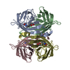 1sweS S: Starting model for refinement C: citing same article ( |
|---|---|
| Similar structure data |

-Links
 PDBj
PDBj- Assembly
| Deposited unit | 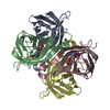
| ||||||||
|---|---|---|---|---|---|---|---|---|---|
| 1 |
| ||||||||
| Unit cell |
|
-Components
| #1: Protein | Mass: 13221.326 Da / Num. of mol.: 4 / Fragment: Core streptavidin (residues 13-139) / Mutation: S45A, D128A Source method: isolated from a genetically manipulated source Source: (gene. exp.) Streptomyces avidinii (bacteria) / Gene: core streptavidin / Species (production host): Escherichia coli / Production host: #2: Chemical | ChemComp-BTN /   Mass: 244.311 Da / Num. of mol.: 4 / Source method: obtained synthetically / Formula: C10H16N2O3S Mass: 244.311 Da / Num. of mol.: 4 / Source method: obtained synthetically / Formula: C10H16N2O3S#3: Water | ChemComp-HOH / |  Mass: 18.015 Da / Num. of mol.: 293 / Source method: isolated from a natural source / Formula: H2O Mass: 18.015 Da / Num. of mol.: 293 / Source method: isolated from a natural source / Formula: H2O |
|---|
-Experimental details
-Experiment
| Experiment | Method: X-RAY DIFFRACTION / Number of used crystals: 1 |
|---|
- Sample preparation
| Crystal | Density Matthews: 1.87 Å3/Da / Density % sol: 45.61 % | ||||||||||||||||||||||||
|---|---|---|---|---|---|---|---|---|---|---|---|---|---|---|---|---|---|---|---|---|---|---|---|---|---|
| Crystal grow | Temperature: 294 K / Method: vapor diffusion, sitting drop / pH: 6.5 Details: sodium citrate, cacodylate, pH 6.5, VAPOR DIFFUSION, SITTING DROP, temperature 294K | ||||||||||||||||||||||||
| Crystal grow | *PLUS Method: vapor diffusion, sitting drop | ||||||||||||||||||||||||
| Components of the solutions | *PLUS
|
-Data collection
| Diffraction | Mean temperature: 100 K |
|---|---|
| Diffraction source | Source: SYNCHROTRON / Site: SSRL  / Beamline: BL9-1 / Wavelength: 0.97 / Wavelength: 0.97 Å / Beamline: BL9-1 / Wavelength: 0.97 / Wavelength: 0.97 Å |
| Detector | Type: MARRESEARCH / Detector: IMAGE PLATE / Date: Apr 1, 2001 |
| Radiation | Protocol: SINGLE WAVELENGTH / Monochromatic (M) / Laue (L): M / Scattering type: x-ray |
| Radiation wavelength | Wavelength: 0.97 Å / Relative weight: 1 |
| Reflection | Resolution: 1.65→50 Å / Num. all: 57334 / Num. obs: 57334 / % possible obs: 99.6 % / Observed criterion σ(I): -3 / Rmerge(I) obs: 0.099 / Net I/σ(I): 14.8 |
| Reflection shell | Resolution: 1.65→1.68 Å / Rmerge(I) obs: 0.542 / Mean I/σ(I) obs: 1.5 / % possible all: 98.5 |
| Reflection | *PLUS Num. measured all: 1009902 |
| Reflection shell | *PLUS % possible obs: 98.5 % |
- Processing
| Software |
| |||||||||||||||||||||||||||||||||
|---|---|---|---|---|---|---|---|---|---|---|---|---|---|---|---|---|---|---|---|---|---|---|---|---|---|---|---|---|---|---|---|---|---|---|
| Refinement | Method to determine structure: ISOMORPHOUS Starting model: PDB entry 1SWE Resolution: 1.65→10 Å / Num. parameters: 35047 / Num. restraintsaints: 44145 / Cross valid method: THROUGHOUT / σ(F): 0 / Stereochemistry target values: ENGH AND HUBER / Details: ANISOTROPIC REFINEMENT REDUCED FREE R (NO CUTOFF)
| |||||||||||||||||||||||||||||||||
| Refine analyze | Num. disordered residues: 8 / Occupancy sum hydrogen: 0 / Occupancy sum non hydrogen: 3881 | |||||||||||||||||||||||||||||||||
| Refinement step | Cycle: LAST / Resolution: 1.65→10 Å
| |||||||||||||||||||||||||||||||||
| Refine LS restraints |
| |||||||||||||||||||||||||||||||||
| Software | *PLUS Name: SHELXL / Version: 97 / Classification: refinement | |||||||||||||||||||||||||||||||||
| Refinement | *PLUS Lowest resolution: 10 Å / Rfactor Rfree: 0.295 / Rfactor Rwork: 0.204 | |||||||||||||||||||||||||||||||||
| Solvent computation | *PLUS | |||||||||||||||||||||||||||||||||
| Displacement parameters | *PLUS |
