 Movie
Movie Controller


+ Open data

- Basic information
| Entry | Database: PDB / ID: 6vjk | ||||||
|---|---|---|---|---|---|---|---|
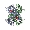
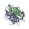
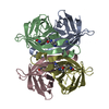
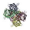
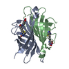
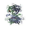
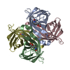
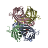
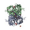
| Title | Streptavidin mutant M88 (N49C/A86C) | ||||||
 Components Components | Streptavidin | ||||||
 Keywords Keywords | Biotin-binding protein / ANTIMICROBIAL PROTEIN | ||||||
| Function / homology |  Function and homology information Function and homology information | ||||||
| Biological species |  Streptomyces avidinii (bacteria) Streptomyces avidinii (bacteria) | ||||||
| Method |  X-RAY DIFFRACTION / SYNCHROTRON / MOLECULAR REPLACEMENT / Resolution: 1.6 Å X-RAY DIFFRACTION / SYNCHROTRON / MOLECULAR REPLACEMENT / Resolution: 1.6 Å | ||||||
 Authors Authors | Marangoni, J.M. / Wu, S.C. / Fogen, D. / Wong, S.L. / Ng, K.K.S. | ||||||
| Funding support |  Canada, 1items Canada, 1items
| ||||||
 Citation Citation | Journal: Sci Rep / Year: 2020 Title: Engineering a disulfide-gated switch in streptavidin enables reversible binding without sacrificing binding affinity. Authors: Marangoni, J.M. / Wu, S.C. / Fogen, D. / Wong, S.L. / Ng, K.K.S. | ||||||
| History |
|
- Structure visualization
| Structure viewer | Molecule: MolmilJmol/JSmol |
|---|
- Downloads & links
-Download
| PDBx/mmCIF format | 6vjk.cif.gz | 325.7 KB | Display | PDBx/mmCIF format |
|---|---|---|---|---|
| PDB format | pdb6vjk.ent.gz | 255.3 KB | Display | PDB format |
| PDBx/mmJSON format | 6vjk.json.gz | Tree view | PDBx/mmJSON format | |
| Others |  Other downloads Other downloads |
-Validation report
| Arichive directory | https://data.pdbj.org/pub/pdb/validation_reports/vj/6vjkftp://data.pdbj.org/pub/pdb/validation_reports/vj/6vjk | HTTPS FTP |
|---|
-Related structure data
| Related structure data | 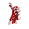 6vjlC 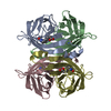 1sweS S: Starting model for refinement C: citing same article ( |
|---|---|
| Similar structure data |

-Links
 PDBj
PDBj- Assembly
| Deposited unit | 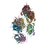
| ||||||||
|---|---|---|---|---|---|---|---|---|---|
| 1 | 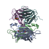
| ||||||||
| 2 | 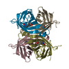
| ||||||||
| 3 | 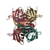
| ||||||||
| Unit cell |
|
-Components
| #1: Protein | Mass: 13133.327 Da / Num. of mol.: 12 / Mutation: N49C, A86C Source method: isolated from a genetically manipulated source Source: (gene. exp.) Streptomyces avidinii (bacteria) / Plasmid: pET29B / Production host: #2: Chemical | ChemComp-BTN /   Mass: 244.311 Da / Num. of mol.: 12 / Source method: obtained synthetically / Formula: C10H16N2O3S / Feature type: SUBJECT OF INVESTIGATION Mass: 244.311 Da / Num. of mol.: 12 / Source method: obtained synthetically / Formula: C10H16N2O3S / Feature type: SUBJECT OF INVESTIGATION#3: Water | ChemComp-HOH / |  Mass: 18.015 Da / Num. of mol.: 1667 / Source method: isolated from a natural source / Formula: H2O Mass: 18.015 Da / Num. of mol.: 1667 / Source method: isolated from a natural source / Formula: H2OHas ligand of interest | Y | Has protein modification | Y | |
|---|
-Experimental details
-Experiment
| Experiment | Method: X-RAY DIFFRACTION / Number of used crystals: 1 |
|---|
- Sample preparation
| Crystal | Density Matthews: 2.2 Å3/Da / Density % sol: 44.05 % |
|---|---|
| Crystal grow | Temperature: 298 K / Method: vapor diffusion, hanging drop / pH: 7.5 Details: 21% PEG 3350, 8% glycerol, 100 mM Bis-Tris-Cl pH 7.5 |
-Data collection
| Diffraction | Mean temperature: 100 K / Serial crystal experiment: N |
|---|---|
| Diffraction source | Source: SYNCHROTRON / Site: SSRL  / Beamline: BL12-2 / Wavelength: 0.98 Å / Beamline: BL12-2 / Wavelength: 0.98 Å |
| Detector | Type: DECTRIS PILATUS 300K / Detector: PIXEL / Date: Apr 10, 2018 |
| Radiation | Protocol: SINGLE WAVELENGTH / Monochromatic (M) / Laue (L): M / Scattering type: x-ray |
| Radiation wavelength | Wavelength: 0.98 Å / Relative weight: 1 |
| Reflection | Resolution: 1.6→50.0005 Å / Num. obs: 176700 / % possible obs: 98.7 % / Redundancy: 8.1 % / Biso Wilson estimate: 16.2 Å2 / CC1/2: 0.994 / Rmerge(I) obs: 0.133 / Rpim(I) all: 0.048 / Rrim(I) all: 0.142 / Rsym value: 0.133 / Net I/σ(I): 13 |
| Reflection shell | Resolution: 1.6→1.69 Å / Redundancy: 8.1 % / Rmerge(I) obs: 0.747 / Mean I/σ(I) obs: 5.4 / Num. unique obs: 25745 / CC1/2: 0.814 / Rpim(I) all: 0.262 / Rrim(I) all: 0.793 / Rsym value: 0.747 / % possible all: 99.4 |
- Processing
| Software |
| ||||||||||||||||||||||||||||||||||||||||||||||||||||||||||||||||||||||||||||||||||||||||||||||||||||||||||||||||||||||||||||||||||||||||||||||||||||||||||||||||||||||||
|---|---|---|---|---|---|---|---|---|---|---|---|---|---|---|---|---|---|---|---|---|---|---|---|---|---|---|---|---|---|---|---|---|---|---|---|---|---|---|---|---|---|---|---|---|---|---|---|---|---|---|---|---|---|---|---|---|---|---|---|---|---|---|---|---|---|---|---|---|---|---|---|---|---|---|---|---|---|---|---|---|---|---|---|---|---|---|---|---|---|---|---|---|---|---|---|---|---|---|---|---|---|---|---|---|---|---|---|---|---|---|---|---|---|---|---|---|---|---|---|---|---|---|---|---|---|---|---|---|---|---|---|---|---|---|---|---|---|---|---|---|---|---|---|---|---|---|---|---|---|---|---|---|---|---|---|---|---|---|---|---|---|---|---|---|---|---|---|---|---|
| Refinement | Method to determine structure: MOLECULAR REPLACEMENT Starting model: 1SWE Resolution: 1.6→50 Å / Cor.coef. Fo:Fc: 0.958 / Cor.coef. Fo:Fc free: 0.943 / SU B: 1.644 / SU ML: 0.059 / Cross valid method: FREE R-VALUE / ESU R: 0.099 / ESU R Free: 0.097
| ||||||||||||||||||||||||||||||||||||||||||||||||||||||||||||||||||||||||||||||||||||||||||||||||||||||||||||||||||||||||||||||||||||||||||||||||||||||||||||||||||||||||
| Solvent computation | Ion probe radii: 0.8 Å / Shrinkage radii: 0.8 Å / VDW probe radii: 1.4 Å | ||||||||||||||||||||||||||||||||||||||||||||||||||||||||||||||||||||||||||||||||||||||||||||||||||||||||||||||||||||||||||||||||||||||||||||||||||||||||||||||||||||||||
| Displacement parameters | Biso mean: 20.982 Å2
| ||||||||||||||||||||||||||||||||||||||||||||||||||||||||||||||||||||||||||||||||||||||||||||||||||||||||||||||||||||||||||||||||||||||||||||||||||||||||||||||||||||||||
| Refinement step | Cycle: LAST / Resolution: 1.6→50 Å
| ||||||||||||||||||||||||||||||||||||||||||||||||||||||||||||||||||||||||||||||||||||||||||||||||||||||||||||||||||||||||||||||||||||||||||||||||||||||||||||||||||||||||
| Refine LS restraints |
| ||||||||||||||||||||||||||||||||||||||||||||||||||||||||||||||||||||||||||||||||||||||||||||||||||||||||||||||||||||||||||||||||||||||||||||||||||||||||||||||||||||||||
| LS refinement shell | Refine-ID: X-RAY DIFFRACTION / Total num. of bins used: 20
|
