 Movie
Movie Controller


+ Open data

- Basic information
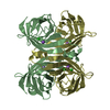
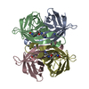
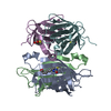
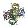
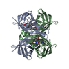
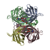
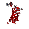
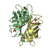
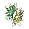
| Entry | Database: PDB / ID: 1i9h | ||||||
|---|---|---|---|---|---|---|---|
| Title | CORE STREPTAVIDIN-BNA COMPLEX | ||||||
 Components Components | STREPTAVIDIN | ||||||
 Keywords Keywords | UNKNOWN FUNCTION / classical beta barrel / protein-ligand complex | ||||||
| Function / homology |  Function and homology information Function and homology information | ||||||
| Biological species |  Streptomyces avidinii (bacteria) Streptomyces avidinii (bacteria) | ||||||
| Method |  X-RAY DIFFRACTION / MOLECULAR REPLACEMENT / Resolution: 2.4 Å X-RAY DIFFRACTION / MOLECULAR REPLACEMENT / Resolution: 2.4 Å | ||||||
 Authors Authors | Livnah, O. / Huberman, T. / Wilchek, M. / Bayer, E.A. / Eisenberg-Domovich, Y. | ||||||
 Citation Citation | Journal: J.Biol.Chem. / Year: 2001 Title: Chicken avidin exhibits pseudo-catalytic properties. Biochemical, structural, and electrostatic consequences. Authors: Huberman, T. / Eisenberg-Domovich, Y. / Gitlin, G. / Kulik, T. / Bayer, E.A. / Wilchek, M. / Livnah, O. | ||||||
| History |
|
- Structure visualization
| Structure viewer | Molecule: MolmilJmol/JSmol |
|---|
- Downloads & links
-Download
| PDBx/mmCIF format | 1i9h.cif.gz | 61.2 KB | Display | PDBx/mmCIF format |
|---|---|---|---|---|
| PDB format | pdb1i9h.ent.gz | 44.8 KB | Display | PDB format |
| PDBx/mmJSON format | 1i9h.json.gz | Tree view | PDBx/mmJSON format | |
| Others |  Other downloads Other downloads |
-Validation report
| Arichive directory | https://data.pdbj.org/pub/pdb/validation_reports/i9/1i9hftp://data.pdbj.org/pub/pdb/validation_reports/i9/1i9h | HTTPS FTP |
|---|
-Related structure data
| Related structure data | 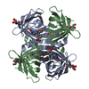 1ij8C  2izfS S: Starting model for refinement C: citing same article ( |
|---|---|
| Similar structure data |

-Links
 PDBj
PDBj- Assembly
| Deposited unit | 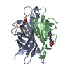
| ||||||||
|---|---|---|---|---|---|---|---|---|---|
| 1 | 
| ||||||||
| Unit cell |
|
-Components
| #1: Protein | Mass: 14497.639 Da / Num. of mol.: 2 / Fragment: RESIDUES 25-163 Source method: isolated from a genetically manipulated source Source: (gene. exp.) Streptomyces avidinii (bacteria) / Production host: #2: Chemical | 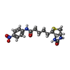  Mass: 364.419 Da / Num. of mol.: 2 / Source method: obtained synthetically / Formula: C16H20N4O4S Mass: 364.419 Da / Num. of mol.: 2 / Source method: obtained synthetically / Formula: C16H20N4O4S#3: Water | ChemComp-HOH / |  Mass: 18.015 Da / Num. of mol.: 134 / Source method: isolated from a natural source / Formula: H2O Mass: 18.015 Da / Num. of mol.: 134 / Source method: isolated from a natural source / Formula: H2O |
|---|
-Experimental details
-Experiment
| Experiment | Method: X-RAY DIFFRACTION / Number of used crystals: 1 |
|---|
- Sample preparation
| Crystal | Density Matthews: 2.04 Å3/Da / Density % sol: 39.56 % | ||||||||||||||||||||
|---|---|---|---|---|---|---|---|---|---|---|---|---|---|---|---|---|---|---|---|---|---|
| Crystal grow | Temperature: 293 K / Method: vapor diffusion, hanging drop / pH: 7.5 Details: 10% isopropanol, 0.1M NaCitrate, 0.05M HEPES, pH 7.5, VAPOR DIFFUSION, HANGING DROP, temperature 293K | ||||||||||||||||||||
| Crystal grow | *PLUS | ||||||||||||||||||||
| Components of the solutions | *PLUS
|
-Data collection
| Diffraction | Mean temperature: 293 K |
|---|---|
| Diffraction source | Source: ROTATING ANODE / Type: RIGAKU RU300 / Wavelength: 1.54 Å |
| Detector | Type: RIGAKU RAXIS IIC / Detector: IMAGE PLATE / Date: Jun 20, 1998 / Details: short Supper Mirrors |
| Radiation | Monochromator: Chales Supper short mirrors / Protocol: SINGLE WAVELENGTH / Monochromatic (M) / Laue (L): M / Scattering type: x-ray |
| Radiation wavelength | Wavelength: 1.54 Å / Relative weight: 1 |
| Reflection | Resolution: 2.4→50 Å / Num. all: 8647 / Num. obs: 8647 / % possible obs: 90.7 % / Observed criterion σ(F): 1 / Observed criterion σ(I): 1 / Redundancy: 2 % / Biso Wilson estimate: 15.3 Å2 / Rmerge(I) obs: 0.098 / Net I/σ(I): 10.9 |
| Reflection shell | Resolution: 2.4→2.5 Å / Redundancy: 2 % / Rmerge(I) obs: 0.265 / % possible all: 94.2 |
| Reflection | *PLUS Lowest resolution: 50 Å / Redundancy: 2 % |
| Reflection shell | *PLUS % possible obs: 94.2 % / Mean I/σ(I) obs: 3.7 |
- Processing
| Software |
| ||||||||||||||||||||||||||||||||
|---|---|---|---|---|---|---|---|---|---|---|---|---|---|---|---|---|---|---|---|---|---|---|---|---|---|---|---|---|---|---|---|---|---|
| Refinement | Method to determine structure: MOLECULAR REPLACEMENT Starting model: 2IZF Resolution: 2.4→50 Å / Cross valid method: THROUGHOUT / σ(F): 1 / σ(I): 0
| ||||||||||||||||||||||||||||||||
| Solvent computation | ksol: 0.335388 e/Å3 | ||||||||||||||||||||||||||||||||
| Displacement parameters |
| ||||||||||||||||||||||||||||||||
| Refinement step | Cycle: LAST / Resolution: 2.4→50 Å
| ||||||||||||||||||||||||||||||||
| Refine LS restraints |
| ||||||||||||||||||||||||||||||||
| Software | *PLUS Name: CNS / Classification: refinement | ||||||||||||||||||||||||||||||||
| Refinement | *PLUS Highest resolution: 2.4 Å / Lowest resolution: 50 Å / σ(F): 1 / % reflection Rfree: 5 % / Rfactor obs: 0.177 | ||||||||||||||||||||||||||||||||
| Solvent computation | *PLUS | ||||||||||||||||||||||||||||||||
| Displacement parameters | *PLUS | ||||||||||||||||||||||||||||||||
| Refine LS restraints | *PLUS
|
