 Movie
Movie Controller


+ Open data

- Basic information
| Entry | Database: PDB / ID: 1swp | ||||||
|---|---|---|---|---|---|---|---|
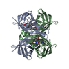
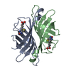
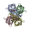
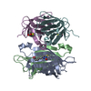
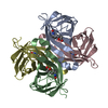
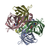
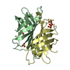
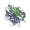
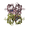
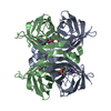
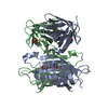
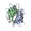
| Title | CORE-STREPTAVIDIN MUTANT W120F IN COMPLEX WITH BIOTIN AT PH 7.5 | ||||||
 Components Components | CORE-STREPTAVIDIN | ||||||
 Keywords Keywords | BIOTIN-BINDING PROTEIN | ||||||
| Function / homology |  Function and homology information Function and homology information | ||||||
| Biological species |  Streptomyces avidinii (bacteria) Streptomyces avidinii (bacteria) | ||||||
| Method |  X-RAY DIFFRACTION / MOLECULAR REPLACEMENT / Resolution: 2 Å X-RAY DIFFRACTION / MOLECULAR REPLACEMENT / Resolution: 2 Å | ||||||
 Authors Authors | Freitag, S. / Le Trong, I. / Chilkoti, A. / Klumb, L.A. / Stayton, P.S. / Stenkamp, R.E. | ||||||
 Citation Citation | Journal: J.Mol.Biol. / Year: 1998 Title: Structural studies of binding site tryptophan mutants in the high-affinity streptavidin-biotin complex. Authors: Freitag, S. / Le Trong, I. / Chilkoti, A. / Klumb, L.A. / Stayton, P.S. / Stenkamp, R.E. #1: Journal: Protein Sci. / Year: 1998Title: Thermodynamic and Structural Consequences of Flexible Loop Deletion by Circular Permutation in the Streptavidin-Biotin System Authors: Chu, V. / Freitag, S. / Le Trong, I. / Stenkamp, R.E. / Stayton, P.S. #2: Journal: Protein Sci. / Year: 1997Title: Structural Studies of the Streptavidin Binding Loop Authors: Freitag, S. / Le Trong, I. / Klumb, L. / Stayton, P.S. / Stenkamp, R.E. | ||||||
| History |
|
- Structure visualization
| Structure viewer | Molecule: MolmilJmol/JSmol |
|---|
- Downloads & links
-Download
| PDBx/mmCIF format | 1swp.cif.gz | 103.6 KB | Display | PDBx/mmCIF format |
|---|---|---|---|---|
| PDB format | pdb1swp.ent.gz | 79.8 KB | Display | PDB format |
| PDBx/mmJSON format | 1swp.json.gz | Tree view | PDBx/mmJSON format | |
| Others |  Other downloads Other downloads |
-Validation report
| Arichive directory | https://data.pdbj.org/pub/pdb/validation_reports/sw/1swpftp://data.pdbj.org/pub/pdb/validation_reports/sw/1swp | HTTPS FTP |
|---|
-Related structure data
| Related structure data | 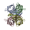 1swhC 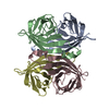 1swjC 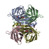 1swkC  1swlC 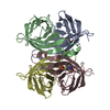 1swnC  1swoC 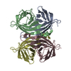 1swqC  1swrC 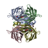 1sweS S: Starting model for refinement C: citing same article ( |
|---|---|
| Similar structure data |
-Links
 PDBj
PDBj- Assembly
| Deposited unit | 
| ||||||||||||||||||||||||||||
|---|---|---|---|---|---|---|---|---|---|---|---|---|---|---|---|---|---|---|---|---|---|---|---|---|---|---|---|---|---|
| 1 |
| ||||||||||||||||||||||||||||
| Unit cell |
| ||||||||||||||||||||||||||||
| Noncrystallographic symmetry (NCS) | NCS oper:
|
-Components
| #1: Protein | Mass: 13242.301 Da / Num. of mol.: 4 / Mutation: W120F Source method: isolated from a genetically manipulated source Source: (gene. exp.) Streptomyces avidinii (bacteria) / Description: PET-210, NOVAGEN, INC., MADISON,WI / Plasmid: PET-210 / Production host: #2: Chemical |   Mass: 244.311 Da / Num. of mol.: 3 / Source method: obtained synthetically / Formula: C10H16N2O3S Mass: 244.311 Da / Num. of mol.: 3 / Source method: obtained synthetically / Formula: C10H16N2O3S#3: Chemical | ChemComp-BTQ / |   Mass: 244.311 Da / Num. of mol.: 1 / Source method: obtained synthetically / Formula: C10H16N2O3S Mass: 244.311 Da / Num. of mol.: 1 / Source method: obtained synthetically / Formula: C10H16N2O3S#4: Water | ChemComp-HOH / |  Mass: 18.015 Da / Num. of mol.: 185 / Source method: isolated from a natural source / Formula: H2O Mass: 18.015 Da / Num. of mol.: 185 / Source method: isolated from a natural source / Formula: H2O |
|---|
-Experimental details
-Experiment
| Experiment | Method: X-RAY DIFFRACTION / Number of used crystals: 1 |
|---|
- Sample preparation
| Crystal | Density Matthews: 2.43 Å3/Da / Density % sol: 49 % | ||||||||||||||||||||||||||||||
|---|---|---|---|---|---|---|---|---|---|---|---|---|---|---|---|---|---|---|---|---|---|---|---|---|---|---|---|---|---|---|---|
| Crystal grow | pH: 7.5 / Details: pH 7.5 | ||||||||||||||||||||||||||||||
| Crystal grow | *PLUS Method: vapor diffusion | ||||||||||||||||||||||||||||||
| Components of the solutions | *PLUS
|
-Data collection
| Diffraction | Mean temperature: 295 K |
|---|---|
| Diffraction source | Source: ROTATING ANODE / Type: RIGAKU RUH2R / Wavelength: 1.5418 |
| Detector | Type: SIEMENS / Detector: AREA DETECTOR / Date: Nov 13, 1996 |
| Radiation | Monochromator: GRAPHITE(002) / Monochromatic (M) / Laue (L): M / Scattering type: x-ray |
| Radiation wavelength | Wavelength: 1.5418 Å / Relative weight: 1 |
| Reflection | Highest resolution: 2.2 Å / Num. obs: 24596 / % possible obs: 90 % / Observed criterion σ(I): 1 / Rmerge(I) obs: 0.07 / Net I/σ(I): 6.9 |
| Reflection shell | Resolution: 2.2→2.3 Å / Rmerge(I) obs: 0.35 / Mean I/σ(I) obs: 1.5 / % possible all: 75 |
| Reflection | *PLUS Num. measured all: 62223 |
| Reflection shell | *PLUS % possible obs: 75 % |
- Processing
| Software |
| |||||||||||||||||||||||||||||||||
|---|---|---|---|---|---|---|---|---|---|---|---|---|---|---|---|---|---|---|---|---|---|---|---|---|---|---|---|---|---|---|---|---|---|---|
| Refinement | Method to determine structure: MOLECULAR REPLACEMENT Starting model: PDB ENTRY 1SWE Resolution: 2→10 Å / Num. parameters: 14951 / Num. restraintsaints: 27450 / Cross valid method: RFREE / σ(F): 0 / Stereochemistry target values: ENGH AND HUBER Details: NON CRYSTALLOGRAPHIC SYMMETRY RESTRAINTS WERE APPLIED FOR THE FOUR SUBUNITS OF THE TETRAMER
| |||||||||||||||||||||||||||||||||
| Solvent computation | Solvent model: MOEWS & KRETSINGER, J.MOL.BIOL.91(1973)201-2 | |||||||||||||||||||||||||||||||||
| Refine analyze | Num. disordered residues: 0 / Occupancy sum hydrogen: 3264 / Occupancy sum non hydrogen: 3737 | |||||||||||||||||||||||||||||||||
| Refinement step | Cycle: LAST / Resolution: 2→10 Å
| |||||||||||||||||||||||||||||||||
| Refine LS restraints |
| |||||||||||||||||||||||||||||||||
| Software | *PLUS Name: SHELXL-97 / Classification: refinement | |||||||||||||||||||||||||||||||||
| Refinement | *PLUS Num. reflection all: 24596 / Rfactor all: 0.202 / Rfactor Rfree: 0.278 | |||||||||||||||||||||||||||||||||
| Solvent computation | *PLUS | |||||||||||||||||||||||||||||||||
| Displacement parameters | *PLUS | |||||||||||||||||||||||||||||||||
| Refine LS restraints | *PLUS Type: s_plane_restr / Dev ideal: 0.015 |
