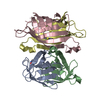
 Movie
Movie Controller


+ Open data

- Basic information
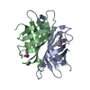
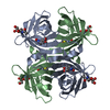
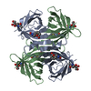
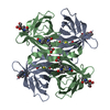
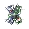
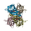
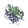
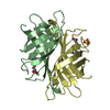
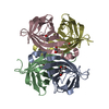
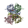
| Entry | Database: PDB / ID: 1lcv | ||||||
|---|---|---|---|---|---|---|---|
| Title | streptavidin-norbiotin complex | ||||||
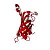
 Components Components | streptavidin | ||||||
 Keywords Keywords | UNKNOWN FUNCTION / avidin / streptavidin / biotin / biotin-analogues | ||||||
| Function / homology |  Function and homology information Function and homology information | ||||||
| Biological species |  Streptomyces avidinii (bacteria) Streptomyces avidinii (bacteria) | ||||||
| Method |  X-RAY DIFFRACTION / MOLECULAR REPLACEMENT / Resolution: 2.3 Å X-RAY DIFFRACTION / MOLECULAR REPLACEMENT / Resolution: 2.3 Å | ||||||
 Authors Authors | Livnah, O. / Pazy, Y. | ||||||
 Citation Citation | Journal: J.Biol.Chem. / Year: 2002 Title: Ligand exchange between proteins: exchange of biotin and biotin derivatives between avidin and streptavidin Authors: Pazy, Y. / Kulik, T. / Bayer, E.A. / Wilchek, M. / Livnah, O. | ||||||
| History |
|
- Structure visualization
| Structure viewer | Molecule: MolmilJmol/JSmol |
|---|
- Downloads & links
-Download
| PDBx/mmCIF format | 1lcv.cif.gz | 58.2 KB | Display | PDBx/mmCIF format |
|---|---|---|---|---|
| PDB format | pdb1lcv.ent.gz | 43 KB | Display | PDB format |
| PDBx/mmJSON format | 1lcv.json.gz | Tree view | PDBx/mmJSON format | |
| Others |  Other downloads Other downloads |
-Validation report
| Arichive directory | https://data.pdbj.org/pub/pdb/validation_reports/lc/1lcvftp://data.pdbj.org/pub/pdb/validation_reports/lc/1lcv | HTTPS FTP |
|---|
-Related structure data


-Links
 PDBj
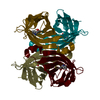
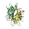
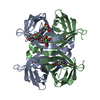
PDBj- Assembly
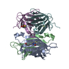
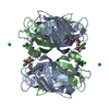
| Deposited unit | 
| ||||||||
|---|---|---|---|---|---|---|---|---|---|
| 1 | 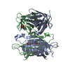
| ||||||||
| 2 | 
| ||||||||
| Unit cell |
| ||||||||
| Details | The second part of the biological assembly is generated by the two fold axis: -x,y -z |
-Components
| #1: Protein | Mass: 12764.833 Da / Num. of mol.: 2 Source method: isolated from a genetically manipulated source Source: (gene. exp.) Streptomyces avidinii (bacteria) / Production host: #2: Chemical | 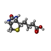  Mass: 230.284 Da / Num. of mol.: 2 / Source method: obtained synthetically / Formula: C9H14N2O3S Mass: 230.284 Da / Num. of mol.: 2 / Source method: obtained synthetically / Formula: C9H14N2O3S#3: Water | ChemComp-HOH / |  Mass: 18.015 Da / Num. of mol.: 94 / Source method: isolated from a natural source / Formula: H2O Mass: 18.015 Da / Num. of mol.: 94 / Source method: isolated from a natural source / Formula: H2O |
|---|
-Experimental details
-Experiment
| Experiment | Method: X-RAY DIFFRACTION / Number of used crystals: 1 |
|---|
- Sample preparation
| Crystal | Density Matthews: 2.32 Å3/Da / Density % sol: 47.06 % | ||||||||||||||||||||||||
|---|---|---|---|---|---|---|---|---|---|---|---|---|---|---|---|---|---|---|---|---|---|---|---|---|---|
| Crystal grow | Temperature: 293 K / Method: vapor diffusion, hanging drop / pH: 4.2 Details: 1.2 M ammonium sulfate, 0.1M sodium acetate, pH 4.2, VAPOR DIFFUSION, HANGING DROP, temperature 293K | ||||||||||||||||||||||||
| Crystal grow | *PLUS Temperature: 20 ℃ / pH: 5.8 | ||||||||||||||||||||||||
| Components of the solutions | *PLUS
|
-Data collection
| Diffraction | Mean temperature: 298 K |
|---|---|
| Diffraction source | Source: ROTATING ANODE / Type: RIGAKU RU300 / Wavelength: 1.5418 |
| Detector | Type: RIGAKU RAXIS IIC / Detector: IMAGE PLATE / Date: Jan 1, 1999 / Details: short supper mirrors |
| Radiation | Monochromator: mirrors / Protocol: SINGLE WAVELENGTH / Monochromatic (M) / Laue (L): M / Scattering type: x-ray |
| Radiation wavelength | Wavelength: 1.5418 Å / Relative weight: 1 |
| Reflection | Resolution: 2.3→40 Å / Num. obs: 10095 / % possible obs: 92.2 % / Observed criterion σ(I): -3 |
| Reflection shell | Resolution: 2.3→2.38 Å / % possible all: 93.4 |
| Reflection | *PLUS Lowest resolution: 50 Å / Redundancy: 2 % / Rmerge(I) obs: 0.094 |
| Reflection shell | *PLUS % possible obs: 93.4 % / Rmerge(I) obs: 0.353 / Mean I/σ(I) obs: 2.3 |
- Processing
| Software |
| ||||||||||||||||||||
|---|---|---|---|---|---|---|---|---|---|---|---|---|---|---|---|---|---|---|---|---|---|
| Refinement | Method to determine structure: MOLECULAR REPLACEMENT / Resolution: 2.3→50 Å / Cross valid method: THROUGHOUT / σ(F): 1 / Stereochemistry target values: Engh & Huber
| ||||||||||||||||||||
| Refinement step | Cycle: LAST / Resolution: 2.3→50 Å
| ||||||||||||||||||||
| Refinement | *PLUS Lowest resolution: 50 Å / % reflection Rfree: 5 % / Rfactor Rfree: 0.234 / Rfactor Rwork: 0.188 | ||||||||||||||||||||
| Solvent computation | *PLUS | ||||||||||||||||||||
| Displacement parameters | *PLUS | ||||||||||||||||||||
| Refine LS restraints | *PLUS
|
