 Movie
Movie Controller


+ Open data

- Basic information
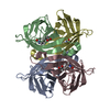
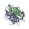
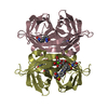
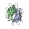
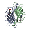
| Entry | Database: PDB / ID: 1df8 | ||||||
|---|---|---|---|---|---|---|---|
| Title | S45A MUTANT OF STREPTAVIDIN IN COMPLEX WITH BIOTIN | ||||||
 Components Components | PROTEIN (STREPTAVIDIN) | ||||||
 Keywords Keywords | BINDING PROTEIN / BIOTIN BINDING PROTEIN / BIOTIN | ||||||
| Function / homology |  Function and homology information Function and homology information | ||||||
| Biological species |  Streptomyces avidinii (bacteria) Streptomyces avidinii (bacteria) | ||||||
| Method |  X-RAY DIFFRACTION / MOLECULAR REPLACEMENT / Resolution: 1.51 Å X-RAY DIFFRACTION / MOLECULAR REPLACEMENT / Resolution: 1.51 Å | ||||||
 Authors Authors | Hyre, D.E. / Le Trong, I. / Freitag, S. / Stenkamp, R.E. / Stayton, P.S. | ||||||
 Citation Citation | Journal: Protein Sci. / Year: 2000 Title: Ser45 plays an important role in managing both the equilibrium and transition state energetics of the streptavidin-biotin system. Authors: Hyre, D.E. / Le Trong, I. / Freitag, S. / Stenkamp, R.E. / Stayton, P.S. | ||||||
| History |
|
- Structure visualization
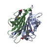
| Structure viewer | Molecule: MolmilJmol/JSmol |
|---|
- Downloads & links
-Download
| PDBx/mmCIF format | 1df8.cif.gz | 66.1 KB | Display | PDBx/mmCIF format |
|---|---|---|---|---|
| PDB format | pdb1df8.ent.gz | 48.1 KB | Display | PDB format |
| PDBx/mmJSON format | 1df8.json.gz | Tree view | PDBx/mmJSON format | |
| Others |  Other downloads Other downloads |
-Validation report
| Arichive directory | https://data.pdbj.org/pub/pdb/validation_reports/df/1df8ftp://data.pdbj.org/pub/pdb/validation_reports/df/1df8 | HTTPS FTP |
|---|
-Related structure data
| Related structure data |  1swaS S: Starting model for refinement |
|---|---|
| Similar structure data |


-Links
 PDBj
PDBj- Assembly
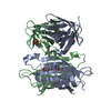
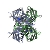
| Deposited unit | 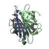
| |||||||||
|---|---|---|---|---|---|---|---|---|---|---|
| 1 | 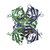
| |||||||||
| 2 | 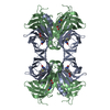
| |||||||||
| Unit cell |
| |||||||||
| Components on special symmetry positions |
|
-Components
| #1: Protein | Mass: 13265.336 Da / Num. of mol.: 2 / Fragment: CORE, RESIDUES 13-139 / Mutation: S45A Source method: isolated from a genetically manipulated source Source: (gene. exp.) Streptomyces avidinii (bacteria) / Production host: #2: Chemical |   Mass: 244.311 Da / Num. of mol.: 2 / Source method: obtained synthetically / Formula: C10H16N2O3S Mass: 244.311 Da / Num. of mol.: 2 / Source method: obtained synthetically / Formula: C10H16N2O3S#3: Water | ChemComp-HOH / |  Mass: 18.015 Da / Num. of mol.: 320 / Source method: isolated from a natural source / Formula: H2O Mass: 18.015 Da / Num. of mol.: 320 / Source method: isolated from a natural source / Formula: H2O |
|---|
-Experimental details
-Experiment
| Experiment | Method: X-RAY DIFFRACTION / Number of used crystals: 1 |
|---|
- Sample preparation
| Crystal | Density Matthews: 1 Å3/Da / Density % sol: 42.46 % | ||||||||||||||||||||
|---|---|---|---|---|---|---|---|---|---|---|---|---|---|---|---|---|---|---|---|---|---|
| Crystal grow | Temperature: 294 K / pH: 4.5 Details: 40% SATURATED AMMONIUM SULFATE, 0.1 M SODIUM ACETATE, pH 4.5, temperature 294.K | ||||||||||||||||||||
| Crystal grow | *PLUS Method: unknown | ||||||||||||||||||||
| Components of the solutions | *PLUS
|
-Data collection
| Diffraction | Mean temperature: 127 K |
|---|---|
| Diffraction source | Source: ROTATING ANODE / Type: RIGAKU RU200 / Wavelength: 1.5418 |
| Detector | Type: RIGAKU / Detector: IMAGE PLATE / Date: Oct 23, 1998 |
| Radiation | Monochromator: GRAPHITE / Protocol: SINGLE WAVELENGTH / Monochromatic (M) / Laue (L): M / Scattering type: x-ray |
| Radiation wavelength | Wavelength: 1.5418 Å / Relative weight: 1 |
| Reflection | Resolution: 1.5→10 Å / Num. all: 167399 / Num. obs: 30427 / % possible obs: 82.4 % / Observed criterion σ(F): 0 / Observed criterion σ(I): 0 / Redundancy: 5.5 % / Rmerge(I) obs: 0.052 / Net I/σ(I): 2.15 |
| Reflection shell | Resolution: 1.5→1.53 Å / Redundancy: 1 % / Rmerge(I) obs: 0.103 / % possible all: 4 |
| Reflection | *PLUS Num. measured all: 167399 |
- Processing
| Software |
| |||||||||||||||||||||||||||||||||
|---|---|---|---|---|---|---|---|---|---|---|---|---|---|---|---|---|---|---|---|---|---|---|---|---|---|---|---|---|---|---|---|---|---|---|
| Refinement | Method to determine structure: MOLECULAR REPLACEMENT Starting model: PDB ENTRY 1SWA Resolution: 1.51→10 Å / Num. parameters: 872 / Num. restraintsaints: 795 / Cross valid method: FREE R / σ(F): 0 / σ(I): 0 / Stereochemistry target values: ENGH AND HUBER
| |||||||||||||||||||||||||||||||||
| Refine analyze | Occupancy sum hydrogen: 1695 / Occupancy sum non hydrogen: 2129 | |||||||||||||||||||||||||||||||||
| Refinement step | Cycle: LAST / Resolution: 1.51→10 Å
| |||||||||||||||||||||||||||||||||
| Refine LS restraints |
| |||||||||||||||||||||||||||||||||
| Software | *PLUS Name: SHELXL-97 / Classification: refinement | |||||||||||||||||||||||||||||||||
| Refinement | *PLUS Lowest resolution: 10 Å / Num. reflection all: 30298 / σ(F): 4 / % reflection Rfree: 10 % / Rfactor all: 0.169 / Rfactor obs: 0.165 / Rfactor Rfree: 0.217 / Rfactor Rwork: 0.166 | |||||||||||||||||||||||||||||||||
| Solvent computation | *PLUS | |||||||||||||||||||||||||||||||||
| Displacement parameters | *PLUS | |||||||||||||||||||||||||||||||||
| Refine LS restraints | *PLUS
|
