 Movie
Movie Controller

[English] 日本語
 Yorodumi
- PDB-1kl5: an engineered streptavidin with improved affinity for the strep-t... -
+ Open data

- Basic information
| Entry | Database: PDB / ID: 1kl5 | ||||||
|---|---|---|---|---|---|---|---|
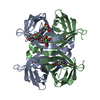
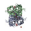
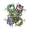
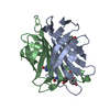
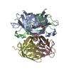
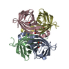
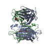
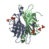
| Title | an engineered streptavidin with improved affinity for the strep-tag II peptide : SAm2-StrepII | ||||||
 Components Components |
| ||||||
 Keywords Keywords | PEPTIDE BINDING PROTEIN / BIOTIN / PROTEIN ENGINEERING / STREP-TAG / STREPTAVIDIN | ||||||
| Function / homology |  Function and homology information Function and homology information | ||||||
| Biological species |  Streptomyces avidinii (bacteria) Streptomyces avidinii (bacteria) | ||||||
| Method |  X-RAY DIFFRACTION / FOURIER SYNTHESIS / Resolution: 1.8 Å X-RAY DIFFRACTION / FOURIER SYNTHESIS / Resolution: 1.8 Å | ||||||
 Authors Authors | Korndoerfer, I.P. / Skerra, A. | ||||||
 Citation Citation | Journal: Protein Sci. / Year: 2002 Title: Improved affinity of engineered streptavidin for the Strep-tag II peptide is due to a fixed open conformation of the lid-like loop at the binding site. Authors: Korndorfer, I.P. / Skerra, A. | ||||||
| History |
|
- Structure visualization
| Structure viewer | Molecule: MolmilJmol/JSmol |
|---|
- Downloads & links
-Download
| PDBx/mmCIF format | 1kl5.cif.gz | 107.5 KB | Display | PDBx/mmCIF format |
|---|---|---|---|---|
| PDB format | pdb1kl5.ent.gz | 83.1 KB | Display | PDB format |
| PDBx/mmJSON format | 1kl5.json.gz | Tree view | PDBx/mmJSON format | |
| Others |  Other downloads Other downloads |
-Validation report
| Arichive directory | https://data.pdbj.org/pub/pdb/validation_reports/kl/1kl5ftp://data.pdbj.org/pub/pdb/validation_reports/kl/1kl5 | HTTPS FTP |
|---|
-Related structure data
| Related structure data |  1kffC  1kl3C  1kl4C 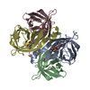 1swuS C: citing same article ( S: Starting model for refinement |
|---|---|
| Similar structure data |


-Links
 PDBj
PDBj- Assembly
| Deposited unit | 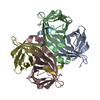
| ||||||||
|---|---|---|---|---|---|---|---|---|---|
| 1 |
| ||||||||
| Unit cell |
| ||||||||
| Details | The biological assembly is one of the teramers in the asymmetric unit |
-Components
| #1: Protein | Mass: 13353.535 Da / Num. of mol.: 4 / Mutation: E44I,S45G,V47R Source method: isolated from a genetically manipulated source Source: (gene. exp.) Streptomyces avidinii (bacteria) / Plasmid: pSA1 / Production host: #2: Protein/peptide | Mass: 1174.263 Da / Num. of mol.: 4 / Source method: obtained synthetically Details: The strep-tag II peptide is chemically synthesized and was selected by synthetic peptide spot assays from a subset of strep-tag derivatives. #3: Water | ChemComp-HOH / |  Mass: 18.015 Da / Num. of mol.: 167 / Source method: isolated from a natural source / Formula: H2O Mass: 18.015 Da / Num. of mol.: 167 / Source method: isolated from a natural source / Formula: H2O |
|---|
-Experimental details
-Experiment
| Experiment | Method: X-RAY DIFFRACTION / Number of used crystals: 1 |
|---|
- Sample preparation
| Crystal | Density Matthews: 2.2 Å3/Da / Density % sol: 44 % | ||||||||||||||||||
|---|---|---|---|---|---|---|---|---|---|---|---|---|---|---|---|---|---|---|---|
| Crystal grow | Temperature: 293 K / Method: vapor diffusion, hanging drop / pH: 7.5 Details: 100 mM Na2HPO4, 1.2-M (NH4)2SO4, pH 7.50, VAPOR DIFFUSION, HANGING DROP, temperature 293K | ||||||||||||||||||
| Crystal grow | *PLUS Temperature: 20 ℃ / pH: 8 | ||||||||||||||||||
| Components of the solutions | *PLUS
|
-Data collection
| Diffraction | Mean temperature: 298 K |
|---|---|
| Diffraction source | Source: ROTATING ANODE / Type: RIGAKU RU300 / Wavelength: 1.5418 |
| Detector | Type: MARRESEARCH / Detector: IMAGE PLATE / Date: Sep 25, 2001 / Details: OSMIC MIRRORS |
| Radiation | Protocol: SINGLE WAVELENGTH / Monochromatic (M) / Laue (L): M / Scattering type: x-ray |
| Radiation wavelength | Wavelength: 1.5418 Å / Relative weight: 1 |
| Reflection | Resolution: 1.8→48 Å / Num. obs: 41071 / % possible obs: 94.5 % / Observed criterion σ(I): -3 / Redundancy: 2.9 % / Rsym value: 0.04 / Net I/σ(I): 19.6 |
| Reflection shell | Resolution: 1.8→1.86 Å / Redundancy: 2.81 % / Mean I/σ(I) obs: 3 / Num. unique all: 3998 / Rsym value: 0.352 / % possible all: 93.2 |
| Reflection | *PLUS Highest resolution: 1.8 Å / Lowest resolution: 99 Å / Num. measured all: 120275 / Rmerge(I) obs: 0.04 |
| Reflection shell | *PLUS % possible obs: 93.2 % |
- Processing
| Software |
| ||||||||||||||||||||
|---|---|---|---|---|---|---|---|---|---|---|---|---|---|---|---|---|---|---|---|---|---|
| Refinement | Method to determine structure: FOURIER SYNTHESIS Starting model: 1SWU Resolution: 1.8→48 Å / SU B: 3.86 / SU ML: 0.122 / σ(F): 0 / ESU R: 0.138 / ESU R Free: 0.132 / Stereochemistry target values: Engh & Huber
| ||||||||||||||||||||
| Displacement parameters | Biso mean: 23.282 Å2
| ||||||||||||||||||||
| Refinement step | Cycle: LAST / Resolution: 1.8→48 Å
| ||||||||||||||||||||
| Refine LS restraints |
| ||||||||||||||||||||
| LS refinement shell | Resolution: 1.8→1.85 Å
| ||||||||||||||||||||
| Refinement | *PLUS Rfactor obs: 0.178 / Rfactor Rfree: 0.221 / Rfactor Rwork: 0.178 | ||||||||||||||||||||
| Solvent computation | *PLUS | ||||||||||||||||||||
| Displacement parameters | *PLUS | ||||||||||||||||||||
| Refine LS restraints | *PLUS
| ||||||||||||||||||||
| LS refinement shell | *PLUS Rfactor Rfree: 0.35 / Rfactor Rwork: 0.28 |
