 Movie
Movie Controller


+ Open data

- Basic information
| Entry | Database: PDB / ID: 1rsu | ||||||
|---|---|---|---|---|---|---|---|
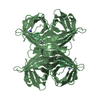
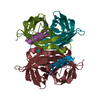
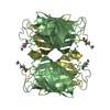
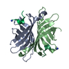
| Title | COMPLEX BETWEEN STREPTAVIDIN AND THE STREP-TAG II PEPTIDE | ||||||
 Components Components |
| ||||||
 Keywords Keywords | COMPLEX (SIGNAL PROTEIN/PEPTIDE) / COMPLEX (SIGNAL PROTEIN-PEPTIDE) / SIGNAL PROTEIN / COMPLEX (SIGNAL PROTEIN-PEPTIDE) complex | ||||||
| Function / homology |  Function and homology information Function and homology information | ||||||
| Biological species |  Streptomyces avidinii (bacteria) Streptomyces avidinii (bacteria) | ||||||
| Method |  X-RAY DIFFRACTION / Resolution: 1.7 Å X-RAY DIFFRACTION / Resolution: 1.7 Å | ||||||
 Authors Authors | Koepke, J. / Schmidt, T. / Skerra, A. | ||||||
 Citation Citation | Journal: J.Mol.Biol. / Year: 1996 Title: Molecular interaction between the Strep-tag affinity peptide and its cognate target, streptavidin. Authors: Schmidt, T.G. / Koepke, J. / Frank, R. / Skerra, A. #1: Journal: J.Chromatogr.,A / Year: 1994Title: One-Step Affinity Purification of Bacterially Produced Proteins by Means of the 'Strep Tag' and Immobilized Recombinant Core Streptavidin Authors: Schmidt, T.G. / Skerra, A. #2: Journal: Protein Eng. / Year: 1993Title: The Random Peptide Library-Assisted Engineering of a C-Terminal Affinity Peptide, Useful for the Detection and Purification of a Functional Ig Fv Fragment Authors: Schmidt, T.G. / Skerra, A. | ||||||
| History |
|
- Structure visualization
| Structure viewer | Molecule: MolmilJmol/JSmol |
|---|
- Downloads & links
-Download
| PDBx/mmCIF format | 1rsu.cif.gz | 48.8 KB | Display | PDBx/mmCIF format |
|---|---|---|---|---|
| PDB format | pdb1rsu.ent.gz | 35.5 KB | Display | PDB format |
| PDBx/mmJSON format | 1rsu.json.gz | Tree view | PDBx/mmJSON format | |
| Others |  Other downloads Other downloads |
-Validation report
| Arichive directory | https://data.pdbj.org/pub/pdb/validation_reports/rs/1rsuftp://data.pdbj.org/pub/pdb/validation_reports/rs/1rsu | HTTPS FTP |
|---|
-Related structure data


-Links
 PDBj
PDBj- Assembly
| Deposited unit | 
| ||||||||||||
|---|---|---|---|---|---|---|---|---|---|---|---|---|---|
| 1 | 
| ||||||||||||
| Unit cell |
| ||||||||||||
| Components on special symmetry positions |
|
-Components
| #1: Protein | Mass: 13341.454 Da / Num. of mol.: 1 / Fragment: RESIDUES 13 - 139 Source method: isolated from a genetically manipulated source Source: (gene. exp.) Streptomyces avidinii (bacteria) / Plasmid: PSA1 / Production host: |
|---|---|
| #2: Protein/peptide | Mass: 1261.341 Da / Num. of mol.: 1 Source method: isolated from a genetically manipulated source |
| #3: Water | ChemComp-HOH /  Mass: 18.015 Da / Num. of mol.: 105 / Source method: isolated from a natural source / Formula: H2O Mass: 18.015 Da / Num. of mol.: 105 / Source method: isolated from a natural source / Formula: H2O |
-Experimental details
-Experiment
| Experiment | Method: X-RAY DIFFRACTION |
|---|
- Sample preparation
| Crystal | Density Matthews: 2.58 Å3/Da / Density % sol: 49 % | ||||||||||||||||||||||||
|---|---|---|---|---|---|---|---|---|---|---|---|---|---|---|---|---|---|---|---|---|---|---|---|---|---|
| Crystal grow | *PLUS pH: 7 / Method: vapor diffusion, hanging drop | ||||||||||||||||||||||||
| Components of the solutions | *PLUS
|
-Data collection
| Diffraction source | Wavelength: 1.5418 |
|---|---|
| Detector | Type: RIGAKU / Detector: IMAGE PLATE / Date: Jun 13, 1994 |
| Radiation | Monochromatic (M) / Laue (L): M / Scattering type: x-ray |
| Radiation wavelength | Wavelength: 1.5418 Å / Relative weight: 1 |
| Reflection | Resolution: 1.7→37.38 Å / Num. obs: 16909 / % possible obs: 97.2 % / Observed criterion σ(I): 0 / Rmerge(I) obs: 0.0923 |
- Processing
| Software |
| ||||||||||||||||||||||||||||||||||||||||||||||||||||||||||||
|---|---|---|---|---|---|---|---|---|---|---|---|---|---|---|---|---|---|---|---|---|---|---|---|---|---|---|---|---|---|---|---|---|---|---|---|---|---|---|---|---|---|---|---|---|---|---|---|---|---|---|---|---|---|---|---|---|---|---|---|---|---|
| Refinement | Resolution: 1.7→8 Å / σ(F): 2
| ||||||||||||||||||||||||||||||||||||||||||||||||||||||||||||
| Displacement parameters | Biso mean: 20.2 Å2 | ||||||||||||||||||||||||||||||||||||||||||||||||||||||||||||
| Refine analyze | Luzzati coordinate error obs: 0.18 Å | ||||||||||||||||||||||||||||||||||||||||||||||||||||||||||||
| Refinement step | Cycle: LAST / Resolution: 1.7→8 Å
| ||||||||||||||||||||||||||||||||||||||||||||||||||||||||||||
| Refine LS restraints |
| ||||||||||||||||||||||||||||||||||||||||||||||||||||||||||||
| Software | *PLUS Name: X-PLOR / Version: 3.1 / Classification: refinement | ||||||||||||||||||||||||||||||||||||||||||||||||||||||||||||
| Refinement | *PLUS Rfactor obs: 0.18 / Rfactor Rwork: 0.18 | ||||||||||||||||||||||||||||||||||||||||||||||||||||||||||||
| Solvent computation | *PLUS | ||||||||||||||||||||||||||||||||||||||||||||||||||||||||||||
| Displacement parameters | *PLUS | ||||||||||||||||||||||||||||||||||||||||||||||||||||||||||||
| Refine LS restraints | *PLUS
|
