 Movie
Movie Controller


+ Open data

- Basic information
| Entry | Database: PDB / ID: 1slg | ||||||
|---|---|---|---|---|---|---|---|
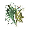
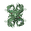
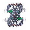
| Title | STREPTAVIDIN, PH 5.6, BOUND TO PEPTIDE FCHPQNT | ||||||
 Components Components |
| ||||||
 Keywords Keywords | COMPLEX(BIOTIN-BINDING PROTEIN/PEPTIDE) / COMPLEX(BIOTIN-BINDING PROTEIN-PEPTIDE) / COMPLEX(BIOTIN-BINDING PROTEIN-PEPTIDE) complex | ||||||
| Function / homology |  Function and homology information Function and homology information | ||||||
| Biological species |  Streptomyces avidinii (bacteria) Streptomyces avidinii (bacteria) | ||||||
| Method |  X-RAY DIFFRACTION / Resolution: 1.76 Å X-RAY DIFFRACTION / Resolution: 1.76 Å | ||||||
 Authors Authors | Katz, B.A. | ||||||
 Citation Citation | Journal: Biochemistry / Year: 1995 Title: Binding to protein targets of peptidic leads discovered by phage display: crystal structures of streptavidin-bound linear and cyclic peptide ligands containing the HPQ sequence Authors: Katz, B.A. | ||||||
| History |
|
- Structure visualization
| Structure viewer | Molecule: MolmilJmol/JSmol |
|---|
- Downloads & links
-Download
| PDBx/mmCIF format | 1slg.cif.gz | 80.9 KB | Display | PDBx/mmCIF format |
|---|---|---|---|---|
| PDB format | pdb1slg.ent.gz | 62 KB | Display | PDB format |
| PDBx/mmJSON format | 1slg.json.gz | Tree view | PDBx/mmJSON format | |
| Others |  Other downloads Other downloads |
-Validation report
| Arichive directory | https://data.pdbj.org/pub/pdb/validation_reports/sl/1slgftp://data.pdbj.org/pub/pdb/validation_reports/sl/1slg | HTTPS FTP |
|---|
-Related structure data


-Links
 PDBj
PDBj- Assembly
| Deposited unit | 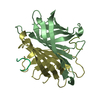
| ||||||||
|---|---|---|---|---|---|---|---|---|---|
| 1 | 
| ||||||||
| Unit cell |
| ||||||||
| Atom site foot note | 1: LYS B 134 - PRO B 135 OMEGA = 148.23 PEPTIDE BOND DEVIATES SIGNIFICANTLY FROM TRANS CONFORMATION | ||||||||
| Noncrystallographic symmetry (NCS) | NCS oper: (Code: given Matrix: (-1, -0.029, -0.004), Vector: Details | MTRIX THE TRANSFORMATIONS PRESENTED ON MTRIX RECORDS BELOW DESCRIBE NON-CRYSTALLOGRAPHIC RELATIONSHIPS AMONG THE VARIOUS DOMAINS IN THIS ENTRY. APPLYING THE APPROPRIATE MTRIX TRANSFORMATION TO THE RESIDUES LISTED FIRST WILL YIELD APPROXIMATE COORDINATES FOR THE RESIDUES LISTED SECOND. NONCRYSTALLOGRAPHIC TWO-FOLD RELATING PROTOMERS OF THE STREPTAVIDIN TETRAMER APPLIED TO TRANSFORMED TO MTRIX RESIDUES RESIDUES RMSD M1 B 13 .. M 7 D 13 .. P 7 0.845 SYMMETRY THE CRYSTALLOGRAPHIC SYMMETRY TRANSFORMATIONS PRESENTED BELOW GENERATE THE SUBUNITS OF THE POLYMERIC MOLECULE. STREPTAVIDIN IS A TETRAMERIC PROTEIN. THE CRYSTALLOGRAPHIC TRANSFORMATION GIVEN HERE GENERATES THE TETRAMER FROM THE DIMER FOUND IN THE ASYMMETRIC UNIT OF THE CRYSTALS. APPLIED TO RESIDUES: B 13 .. B 133 D 13 .. D 133 M 2 .. M 6 P 1 .. P 6 SYMMETRY1 1 1.000000 0.000000 0.000000 0.00000 SYMMETRY2 1 0.000000 -1.000000 0.000000 0.00000 SYMMETRY3 1 0.000000 0.000000 -1.000000 0.00000 | |
-Components
| #1: Protein | Mass: 14181.324 Da / Num. of mol.: 2 / Source method: isolated from a natural source / Source: (natural) Streptomyces avidinii (bacteria) / References: UniProt: P22629#2: Protein/peptide | Mass: 830.864 Da / Num. of mol.: 2 / Source method: isolated from a natural source #3: Water | ChemComp-HOH / |  Mass: 18.015 Da / Num. of mol.: 159 / Source method: isolated from a natural source / Formula: H2O Mass: 18.015 Da / Num. of mol.: 159 / Source method: isolated from a natural source / Formula: H2O |
|---|
-Experimental details
-Experiment
| Experiment | Method: X-RAY DIFFRACTION |
|---|
- Sample preparation
| Crystal | Density Matthews: 2.04 Å3/Da / Density % sol: 39.61 % |
|---|---|
| Crystal grow | pH: 5.6 / Details: pH 5.6 |
| Crystal grow | *PLUS pH: 4 / Method: vapor diffusion, sitting drop |
| Components of the solutions | *PLUS Conc.: 15 mg/ml / Common name: peptide |
-Data collection
| Diffraction source | Wavelength: 1.5418 |
|---|---|
| Detector | Type: SIEMENS / Detector: AREA DETECTOR |
| Radiation | Monochromatic (M) / Laue (L): M / Scattering type: x-ray |
| Radiation wavelength | Wavelength: 1.5418 Å / Relative weight: 1 |
| Reflection | Num. obs: 21587 / Observed criterion σ(I): 3.5 / Redundancy: 4.4 % / Rmerge(I) obs: 0.072 |
| Reflection | *PLUS Highest resolution: 1.74 Å / Lowest resolution: 50 Å / Num. measured all: 94347 / Rmerge(I) obs: 0.072 |
- Processing
| Software |
| ||||||||||||||||||||||||||||||||||||||||||||||||||||||||||||
|---|---|---|---|---|---|---|---|---|---|---|---|---|---|---|---|---|---|---|---|---|---|---|---|---|---|---|---|---|---|---|---|---|---|---|---|---|---|---|---|---|---|---|---|---|---|---|---|---|---|---|---|---|---|---|---|---|---|---|---|---|---|
| Refinement | Resolution: 1.76→7.5 Å / σ(F): 1 Details: CRYST1 CELL AXES CHOSEN TO CORRESPOND TO COORDINATES OF STREPTAVIDIN DEPOSITED BY WEBER ET AL. IN THE PDB (ENTRY 1PTS). THE FOLLOWING ATOMS HAD WEAK DENSITY AND OCCUPANCIES WERE REFINED: B ...Details: CRYST1 CELL AXES CHOSEN TO CORRESPOND TO COORDINATES OF STREPTAVIDIN DEPOSITED BY WEBER ET AL. IN THE PDB (ENTRY 1PTS). THE FOLLOWING ATOMS HAD WEAK DENSITY AND OCCUPANCIES WERE REFINED: B 13, B 14, B 15 AND (NOT NAME C OR NAME O) D 13, D 14, D 15 AND (NOT NAME C OR NAME O) D 46, D 47, D 48, D 49, D 50, D 51 AND (NOT NAME C OR NAME O) B 46, B 47, B 48, B 49, B 50 AND (NOT NAME C OR NAME O) B 67, B 68 AND (NOT NAME C OR NAME O) B 53 AND (NAME NE OR NAME NH1 OR NAME NH2 OR NAME CZ) B 103 AND (NAME NE OR NAME NH1 OR NAME NH2 OR NAME CZ) D 103 AND (NAME NE OR NAME NH1 OR NAME NH2 OR NAME CZ) D 84 AND (NAME NE OR NAME NH1 OR NAME NH2 OR NAME CZ) B 116 AND (NAME CG OR NAME OR NAME OE1 OR NAME OE2) M 7, P 1, P 2, B 135 M 1 WAS NOT LOCATED OR INCLUDED IN THE MODEL. DISCRETELY DISORDERED SIDE CHAINS WHOSE OCCUPANCIES AND STRUCTURES WERE SIMULTANEOUSLY REFINED WERE B 73, D 73, B 110, D 110, B 22, D 107, D 105. B 22 IS DISORDERED BETWEEN 2 CONFORMATIONS ONE OF WHICH OCCUPIES A SIMILAR REGION OF SPACE AS A TWO-FOLD RELATED B 22. THIS DISORDER CAN NOT BE PROPERLY REFINED WITH X-PLOR. SEVERAL WATERS ARE ON OR NEAR TWO-FOLD AXES, AND CAN NOT BE PROPERLY REFINED WITH X-PLOR. BULK SOLVENT WAS REFINED.
| ||||||||||||||||||||||||||||||||||||||||||||||||||||||||||||
| Displacement parameters | Biso mean: 22 Å2 | ||||||||||||||||||||||||||||||||||||||||||||||||||||||||||||
| Refinement step | Cycle: LAST / Resolution: 1.76→7.5 Å
| ||||||||||||||||||||||||||||||||||||||||||||||||||||||||||||
| Refine LS restraints |
| ||||||||||||||||||||||||||||||||||||||||||||||||||||||||||||
| Software | *PLUS Name: X-PLOR / Classification: refinement | ||||||||||||||||||||||||||||||||||||||||||||||||||||||||||||
| Refinement | *PLUS | ||||||||||||||||||||||||||||||||||||||||||||||||||||||||||||
| Solvent computation | *PLUS | ||||||||||||||||||||||||||||||||||||||||||||||||||||||||||||
| Displacement parameters | *PLUS | ||||||||||||||||||||||||||||||||||||||||||||||||||||||||||||
| Refine LS restraints | *PLUS
|
